Unit 2 Basic Immunologic Reactions
Unit 2 Basic Immunologic Reactions. Part 7 Molecular Techniques Terry Kotrla, MS, MT(ASCP)BB. Molecular Diagnostic Assays. Very powerful tools. Quick supplies information to assist in diagnosis and monitoring of diseases. Bacteria Viruses Genetic diseases

Unit 2 Basic Immunologic Reactions
E N D
Presentation Transcript
Unit 2 Basic Immunologic Reactions Part 7 Molecular Techniques Terry Kotrla, MS, MT(ASCP)BB
Molecular Diagnostic Assays • Very powerful tools. • Quick supplies information to assist in diagnosis and monitoring of diseases. • Bacteria • Viruses • Genetic diseases • This technology is impacting every area of the clinical laboratory.
Molecular Techniques • Techniques include • Enzymatic cleavage of nucleic acids • Gel electrophoresis • Enzymatic amplification of target sequences • Hybridization with nucleic acid probes • Advantages and disadvantages of each will be discussed.
Gene • Genes are located on 23 pairs of chromosomes. • DNA is packed into genes. • Genes are units of heredity • DNA organized into long structures called chromosomes, genes are pieces of DNA which correspond to a single unit of inheritance.
Two types of nucleic acids: RNA & DNA • DNA carries genetic information within chromosomes of each cell. • Main role is long-term storage of information. • Blueprint to construct other cell components • RNA • Transcribed from DNA • Central role is protein synthesis
DNA and RNA • DNA is encoded with four interchangeable "building blocks", called "bases", • Adenine and Thymine, which pair together. • Cytosine and Guanine, which pair together. • RNA has five different bases: • Adenine and Uracil, which pair together. • Cytosine and Guanine, which pair together.
Three Main Differences • DNA is double stranded, RNA is single stranded. • DNA contains deoxyribose, RNA contains ribose. • Complementary base to adenine is thymine in DNA and uracil in RNA.
DNA Replication • DNA very stable. • Loses conformational structure under extremities of heat, pH or presence of destablizing agents. • Semi-conservative process, one strand acts as template to create exact copy. • Bonds holding strands together are weak.
DNA Replication – In-Vivo • Strand “unzips”, hydrogen bonds between base pairs are broken. • Sequence of bases on strand serve as template to which complementary bases are added. • When process is complete 2 identical DNA molecules are formed.
DNA Replication – In-Vitro • Two steps • Denaturation – can use heat or alkaline solutions to break bonds and separate the two strands, if heat is used term is called “melting”. • Annealing – strands cool and complementary strands spontaneously rejoin. • This process is exploited in the laboratory.
Types of RNA • One strand of DNA serves as template for messenger or “mRNA”. • mRNA carries information from DNA to ribosome. • Uracil transcribed where Thymine would have occurred. • Transfer RNA (tRNA) transports amino acids to make proteins. • Ribosomal RNA (rRNA) acts as site of protein synthesis directed by mRNA. • RNA less stable and degrades more rapidly than DNA.
Hybridization Techniques • Spontaneous pairing of DNA strands forms the basis for detection and characterization of genes. • Probe technology used to identify individual genes or DNA sequences.
Nucleic Acid Probes • Nucleic acid probe is a short strand of DNA or RNA of known sequence • Used to identify presence of complementary single strand of DNA in patient sample. • Binding of the patient strand with the probe is known as hybridzation.
Nucleic Acid Probes • Two DNA strands must share at least 16 to 20 consecutive bases of perfect complementarity to form stable hybrid. • Match occurring as a result of chance less than 1 in a billion. • Probes labeled with marker: radioisotope, fluorochrome, enzyme or chemiluminescent substrate. • Hybridization can take place in solid support medium or liquid.
Solid Support Hybrization • Dot-blot • Sandwich hybridization • Gel electrophoresis • Southern Blot • Northern Blot
Dot-Blot • Dot-blot clinical sample applied to membrane, heated to denature DNA. • Labeled probes added and will bind to target if present. • Wash to remove unhybridized probe. • Detect by autoradiography or enzyme assay. • Qualitative test only. • May be difficult to interpret.
Sandwich Hybridization • Uses two probes, one bound to membrane and serves as capture target for sample DNA of interest. • Second probe anneals to different site on target DNA and has label for detection. • Sample nucleic acid sandwiched between the two. • Two hybridization events occur, increases specificity. • Can be adapted to microtiter plates.
Characterization of DNA • Restriction endonucleases cleave DNA at SPECIFIC recognition sites approximately 4 to 6 base pairs long. • Enzyme which cuts ds or ss DNA at specific site. • Enzymes found naturally. • Over 3000 identified, 500 available commercially. • Human DNA can yield millions of unique fragments.
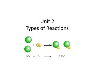
Characterization of DNA • EcoRI popular restriction endonuclease. • Cuts wherever there is a G-C or C-G bond. • Fragments separated based on size and electrical charge by electrophoresis. • Kits developed to detect specific gene fragments associated with diseases or conditions
Characterization of DNA • Differences in restriction patterns referred to as restriction fragment length polymorphisms (RFLP). • Caused by variations in nucleotides within genes that change where restriction enzymes cleave DNA. • When mutations occur different sized pieces of DNA are obtained.
Use of RFLP • Identify specific microorganisms • Detect polymorphism in MHC genes. • DNA fingerprinting – first method used • Localization of genes for genetic disorders • Determination of risk for disease • Paternity testing
Southern Blot • Used to detect a specific DNA sequence in a sample. • Use restriction endonuclease to cut DNA into fragments. • DNA fragments separated by electrophoresis.
Southern Blot • Pieces denatured and transferred from gel to membrane for hybridization reaction. • Place membrane on top of gel, add weights or use suction and allow buffer plus DNA to wick up onto the membrane. • Once DNA is on membrane heat or use UV light to crosslink strands to permanently attach the DNA onto membrane. • Add labeled DNA probes for hybridization to take place. • Radioactive, fluorescent or chromogenic dye used as label • Probes added in excess so target molecules reanneal and more likely to attach to probe. • Visualize results based on label used.
Southern Blot • Southern Blot takes advantage of the fact that DNA fragments will stick to a nylon or nitrocellulose membrane. • Membrane laid on top of the agarose gel and absorbent material (e.g. paper towels or a sponge) is placed on top. • With time, the DNA fragments will travel from the gel to the membrane by capillary action as surrounding liquid is drawn up to the absorbent material on top. • After the transfer of DNA fragments has occurred, the membrane is washed, then the DNA fragments are permanently fixed to the membrane by heating or exposing it to UV light. • The membrane is now a mirror image of the agarose gel.
Southern Blot • http://www.koreanbio.org/Biocourse/index.php/Hybridization
Southern Blot • MOM [blue], DAD [yellow], and their four children: D1 (the biological daughter), D2 (step-daughter, child of Mom and her former husband [red]), S1 (biological son), and S2 (adopted son, not biologically related [his parents are light and dark green]).
Northern Blot • Northern blots used to detect RNA in a sample. • RNA (either total RNA or just mRNA) separated by gel electrophoresis. • RNA transferred to sheet of nitrocellulose, though other membranes can be used and immobilized by heat or UV light. • Blot is incubated with a probe which is single-stranded DNA. • This probe will form base pairs with its complementary RNA sequence and bind to form a double-stranded RNA-DNA molecule. The probe cannot be seen but it is either radioactive or has an enzyme bound to it (e.g. alkaline phosphatase or horseradish peroxidase). • Location of the probe is revealed by incubating with a substrate that the attached enzyme converts to a colored product that can be seen or gives off light when exposed on X-ray film. • If the probe was labeled with radioactivity, it can expose X-ray film directly.
Solution Hybridization • Both target nucleic acid and probe free to interact in solution. • Hybridization of probe to target in solution is more sensitive than hybridization on solid support • Requires less sample and is more sensitive. • Probe must be single-stranded and incapable of self-annealing. • Fairly adaptable to automation, especially those using chemiluminescent labels. • Assays performed in a few hours.
Solution Hybridization • Second method is hybridization protection assay. • Chemiluminescent acridinium ester attached to probe as label. • After hybridization solution subjected to alkaline hydrolysis. • Causes hydrolysis of ester if probe not attached to target. • If probe attaches to target light given off.
In-Situ Hybridization • Target nucleic acid found in intact cells. • Provides information about presence of specific DNA targets and distribution in tissues. • Probes must be small enough to reach nucleic acid. • Radioactive or fluorescent tags used. • Requires experience.
In-Situ Hybridization • Fluorescent in-situ hybridization (FISH) very popular. • Cytogenetic technique used to detect and localize presence or absence of DNA sequences on chromosomes. • Used in genetic counseling, medicine and species identification.
FISH • A metaphase cell positive for the bcr/abl rearrangement (associated with chronic myelogenous leukemia) using FISH. • The chromosomes can be seen in blue. The chromosome that is labeled with green and red spots (upper left) is the one where the wrong rearrangement is present.
FISH • DNA probes specific to regions of particular chromosomes are attached to fluorescent markers and hybridized with a chromosome spread. • Picture shows a computer-generated "false colour" image, in which small variations in fluorescence wavelength among probes are enhanced as distinct primary colours. • The combination of probes that hybridize to a particular chromosome produces a unique pattern for each chromosome. This makes it particularly easy to detect segmental deletions and translocations among chromosomes.
DNA Chip aka Microarrays • A DNA chip (DNA microarray) is a biosensor which analyzes gene information from humans and bacteria. • Utilizes the complementation of the four bases labeled A, T, G and C in which A pairs with T and G pairs with C through hydrogen bonding. • Solution of DNA sequences containing known genes (DNA probe) placed on glass plates in microspots several microns in diameter arranged in multiple rows. • Genes are extracted from samples such as blood, amplified and then reflected in the DNA chip, enabling characteristics such as the presence and mutation of genes in the test subject to be determined. • As gene analysis advances, the field is gaining attention particularly in the clinical diagnosis of infectious disease, cancer and other maladies.
Microarrays • Microarrays are a significant advance both because they may contain a very large number of genes and because of their small size. • Useful in surveying large number of genes quickly or when the sample to be studied is small. • May be used to assay gene expression within a single sample or to compare gene expression in two different cell types or tissue samples, such as in healthy and diseased tissue. • Because a microarray can be used to examine the expression of hundreds or thousands of genes at once, it promises to revolutionize the way scientists examine gene expression. • This technology is still considered to be in its infancy.
Microarray • Fix the single stranded DNA to chip. • Labeled with different colors.
Microarray • A microarray scanner provides a picture of what spots showed up green, red, or yellow. • A green dot would represent a gene that is expressed more in healthy cells or have less expression in cancer cells, and a red dot represents a gene that is expressed more in cancer cells. • A yellow spot would be a gene that is expressed in both cancer and healthy cells.
Drawbacks • Stringency, or correct pairing, is affected by: • Salt concentration • Temperature • Concentration of destabilizing agent such as formamide or urea. • If conditions not carefully controlled mismatches can occur. • Patient nucleic acid may be present in small amounts, below threshold for probe detection. • Sensitivity can be increased by amplification: target, probe and signal
DNA Sequencing • Method to determine the exact order of the nucleotide bases in DNA. • Unknown DNA sequences compared to known. • Several methods available. • Sanger method of choice.
Sanger Method of DNA Sequencing • Requires ss DNA template, DNA primer, DNA polymerase, labeled nucleotides and modified nucleotides to terminate DNA elongation. • DNA sample divided into 4 separate reactions to normal (NTP) and ONE type of dideoxynucleotides (ddNTP) A, T, C or G are added. • ddNTPs will prevent addition of further nucleotides. • Creates DNA strands of discrete sizes. • Each reaction loaded on separate lane on gel and electrophoresed. • Sequence of nucleotides read in order to determine DNA sequence.