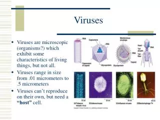

ARBO VIRUSES
E N D
Presentation Transcript
ARBO VIRUSES DR. SUDHEER KHER
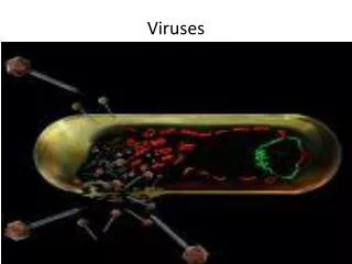
Zoonotic diseases • Zoonotic viruses are viruses which are transmissible from animals (arthropods, vertebrates) to man. Many are transmitted by means of an infected, blood-sucking, arthropod vector (arthropod borne = arboviruses). Others may be transmitted by inhalation, or conjunctival contact with infected excretions, or by direct contact with infected animal (e.g. rabies).
REQUIREMENTS • ARTHROPOD VECTORS • VERTEBRATE HOSTS • DEADEND HOSTS
LIFE CYCLES OF ARBOVIRUSES • There are several types of life cycles, but many arboviruses have a sylvatic cycle while some also have an urban cycle. • 1. Sylvatic cycle (sometimes known as the jungle cycle). In this the virus cycles between an arthropod and a mammalian host with man usually a dead-end host infected by the arthropod • 2. Urban cycle. In this the virus cycles between man and an arthropod species.
ARBOVIRUS DISEASE • Diseases caused by arboviruses include: • encephalitis • febrile diseases (sometimes associated with rash) • hemorrhagic fevers
Dengue: The Problem. • Dengue is an endemic viral disease affecting tropical and subtropical regions around the world, predominantly in urban and semi-urban areas. • Dengue fever (DF) and its more serious forms, dengue hemorrhagic fever (DHF) and dengue shock syndrome (DSS), are becoming important public health problems. • Was formally included within the disease portfolio of the United Nations Development Programme/World Bank/World Health Organization Special Programme for Research and Training in Tropical Diseases by the Joint Coordination Board in June 1999 (WHO, 1999). • The global prevalence of dengue has grown dramatically in recent decades. • The disease is now endemic in more than 100 countries in Africa, the Americas, the eastern Mediterranean, Southeast Asia, and the Western Pacific, threatening more than 2.5 billion people (Gubler, 1998). • The World Health Organization estimates that there may be 50 million to 100 million cases of dengue virus infections worldwide every year, which result in 250,000 to 500,000 cases of DHF and 24,000 deaths each year (WHO, 1997)
Aedes aegypti: - The vector The victim: Mortality can be as high as 10-20% (over 40% if shock occurs.
The break bone fever Dengue Infected cells Dengue Hemorrhage
Dengue Syndrome • An acute viral febrile exanthema, caused by any of the four serotypes (1-4) of the Dengue Viruses, members of Flaviviridae. • Transmitted by mosquitoes of Stegomia family, mainly Aedes aegypti • Dengue virus is a positive-stranded encapsulated RNA virus, with a genome of ~11kb
A broad spectrum of illness is caused, from inapparent infection, flu-like fever and classical DF to DHF-DSS. • No specific treatment, only supportive therapy. • Diagnosis based on clinical syndromes is unreliable. • Laboratory confirmation is needed for: • Timely intervention • Etiologic investigation and • Disease control.
Dengue Case Definition • Patients suspected as dengue cases, presenting with: • High-grade fever (>38.50C), • Mayalgia, • Vomiting, • Hepatomegaly, • Rash - hemorrhagic symptoms. • Tourniquet test +ve, • ESR (>20%) • Low platelet count (<100000/mm3)
Laboratory Diagnosis1. Virus Isolation & Characterization2. Serological Methods3. Molecular Methods (RTPCR)
Serological methods • Antibody Detection: • Indirect Immunofluorescence Antibody Test • Neutralization Test • Hemagglutination Test • Complement Fixation Test • Rapid Immunochromatography TestIgM & IgG ELISA • Antigen Detection: • E/M & NS1 antigen detection through ELISA & Dot-Blot Assays – in acute phase sera upto 9 days after onset.