Download
1 / 11
110 likes | 129 Views
Explore the specialized properties and functions of striated, non-striated skeletal, cardiac, and smooth muscle tissues. Learn about their structure, innervation, and classification. Discover the organization and innervation of skeletal muscles along with the unique features of cardiac and smooth muscles.

E N D
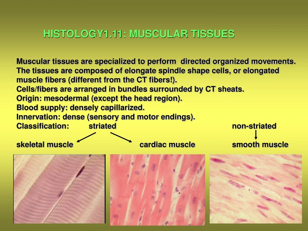
HISTOLOGY1.11: MUSCULAR TISSUES Muscular tissues are specialized to perform directed organized movements. The tissues are composed of elongate spindle shape cells, or elongated muscle fibers (different from the CT fibers!). Cells/fibers are arranged in bundles surrounded by CT sheats. Origin: mesodermal (except the head region). Blood supply: densely capillarized. Innervation: dense (sensory and motor endings). Classification: striated non-striated skeletal muscle cardiac muscle smooth muscle
Skeletal muscle Cardiac muscle Smooth muscle Tissue unit: non-branching Y-shaped cells spindle-shaped multinucleated form fibers cells form fibers fibers Functional quick contraction quick contraction slow contraction properties: easily fatigues no fatigue no fatigue Impulse motor endplate specialized vegetative generation: cardiac muscle plexus Occurrence: locomotor heart wall of visceral organs(muscles) organs
Skeletal muscle Skeletal myofibers vary from 10 to 120 mm. Origin: fusion of many mononuclear myoblast cells. Morphology: 1./Longitudinal section: elongated nuclei at subsarcolemmal position transverse striation as regular cross-banding pattern 2./Cross-section: polygonal-shaped profiles no-cross-striation 1 2
Organization of the muscles: Individual myofibers surrounded by endomysium (fine reticular CT) Primary bundles/fascicles ensheathed by perimysium (dense collagen fibers with blood vessels and nerves) The whole muscle is ensheathed by the epimysium (thick dense collagenous CT)
Sensory innervation of the skeletal muscle: Morphology of the muscle spindle Schematic drawing of the components LM image of muscle spindle
Motor innervation of the skeletal muscle: Light micrograph of the motor endplate Schematic drawing of the Motor endplate
CARDIAC MUSCLE Cardiac myofibers are organized for pumping the blood in the circulatory system. Myofibers are formed by individual cells that branch and anasotomose. Morphology: cylindrical, or Y-shaped cells centrally located single nucleus very dense capillary network cell borders: Eberth’s line (discus intercalaris) (arrows) Longitudinal section Cross-section
CARDIAC MUSCLE Comparison of the cardiac muscle types: The smaller slender fibers with more contractile elements are engaged with contraction, the larger with more sarcoplasm are responsible for impulse generation and conduction Model drawing of the cardiac muscle cells and their attachment to each other by the Eberth’s lines (blue lines)
Cardiac myocytes : Purkinje fibers Isolated cell A bundle with Discus intercalaris formed by capillaries desmosomes (D) and gap junctions (N)
SMOOTH MUSCLE: Smooth muscle myofibers are formed by small mononucleated spindle-shaped cells of 10 mm in diameter. Length varies between 20-500 mm. No cross-striation is visible. No perimysium. Rod-like nucleus is in the center of the cell. Longitudinal section N Cross-section
Organization of smooth muscle cells into fiber bundles and their innervation Contraction of smooth muscle cells