Download
1 / 66
660 likes | 966 Views
Cyanotic Heart Disease; Overview of Management. By Dr. Ahmad Shaker MD Cardiology. Definition.

E N D
Cyanotic Heart Disease;Overview of Management By Dr. Ahmad Shaker MD Cardiology
Definition • Cyanotic Heart Disease is a defect or group of defects in the structure or function of the heart or the great vessels, present at birth, consisting of abnormal blood flow from the right to the left part of the circulatory system (either at the level of the atria, the ventricles, or the great vessels). • This abnormal communication (called right-to-left shunt) results in poor oxygenation of the body and therefore cyanosis
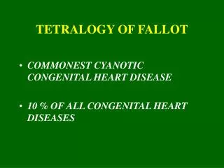
Causes ofCyanotic Heart Disease: • –Tetralogy of Fallot • –Transposition of the great arteries (D-TGA) • –Single ventricle • –Truncus arteriosus • –Total anomalous pulmonary venous connections • –Ebstein’s anomaly • –Eisenmenger’s disease
Ductal Independent Mixing Lesions: -Truncus Arteriosus . -d-Transposition of Great Arteries. -Total Anomalous Pulmonary Venous Connection without Obstruction (TAPVC). Ductal-Dependent Pulmonary Blood flow: -Tricuspid Atresia -Pulmonic Atresia with Intact Ventricular Septum. -Tetralogy of Fallot. -Ebstein’s Anomaly.
Lesions with Ductal Dependent Systemic Blood Flow: -Hypoplastic Left Heart Syndrome (HLHS). -Interrupted Aortic Arch. -Total Anomalous Pulmonary Venous Connection with Obstruction.
Symptoms & Signs • Cyanosis. • Dyspnea. • Failure to thrive, or failure to grow properly • Fatigue • Squatting position after physical activity to relieve breathlessness. • Hypoxic spells, characterized by: -Anxiety. -Hyperventilation. -Sudden increase in cyanosis. • Syncope. • Chest pain, Arrythmias. • CHF.
Diagnosis Clinical: -Upper left sternal border ejection murmur of RV outflow tract obstruction --------- TOF. -Newborns present with severe cyanosis and a continuous murmur of ductal flow --------- pulmonary valve atresia and ductus arteriosus-dependent pulmonary blood flow . -Present immediately after birth with severe cyanosis that progresses rapidly to metabolic acidosis------- TGA.
Days to weeks after birth with heart failure and mild hypoxemia, A hyperdynamic precordium, wide pulse pressure, a normal S1 with a frequent ejection click, and a loud, usually single S2 are characteristic.------ persistant truncus. • In persistent truncus, Heart murmurs vary and may include a flow murmur at the base, a loud regurgitant murmur at the lower left sternal border, and a mid-diastolic mitral flow murmur. With truncal valve insufficiency, a high-pitched diastolic murmur over the mid-sternum is present
-Combined ventricular hypertrophy ----- Truncus. -Right axis deviation and Right Ventricular Hypertrophy ---- Total APVD. -Superior axis and L V Hypertrophy ---- TV atresia. -LV hypertrophy, leftward axis --- PulmonicAtresia with Intact Ventricular Septum -Right Bundle Branch Block, Delta Waves due to WPW syndrome --- Ebestine’s Anomaly.
TGA Right ventricular hypertrophy, right axis deviation .
TFO (Coure in Sabou). • Small heart and a concave main pulmonary artery segment with lung oligemia, Right aortic arch is present in 25%
Cardiomegally with narrow base, lung hyperemia ---- TGA (Egg on side).
A Segmental Approach to cardiac Situs & malposition; 1- Atrial Situs: Veseral Situs & visceroatrial concordance. Atrial morphology (situs solitus or inversus). venous inflow pattern. 2- Ventricular localization: ventricular morphology. atrioventricular concordance. Base-to-apex axis (Levo or Dextrocardia). 3- Greate Artery Connections & its identifications. 4- Ventriculoarterial concordance.