Download
1 / 72
880 likes | 1.42k Views
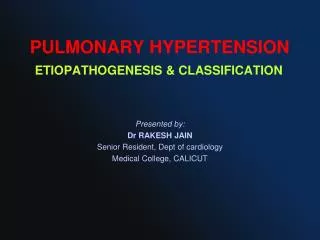
PULMONARY HYPERTENSION ETIOPATHOGENESIS & CLASSIFICATION. Presented by : Dr RAKESH JAIN Senior Resident, Dept of cardiology Medical College, CALICUT. DEFINITION OF PAH. Current hemodynamic definition is a mPAP >25 mm Hg PCWP, LA pressure, or LVEDP ≤15 mm Hg, and PVR>3 Wood units.

E N D
PULMONARY HYPERTENSIONETIOPATHOGENESIS & CLASSIFICATION Presented by: Dr RAKESH JAIN Senior Resident, Dept of cardiology Medical College, CALICUT
DEFINITION OF PAH Current hemodynamic definition is a • mPAP >25 mm Hg • PCWP, LA pressure, or LVEDP ≤15 mm Hg, and • PVR>3 Wood units Circulation. 2009;119:2250-2294 J Am Coll Cardiol.2009;53:1573-1619
WHO Geneva, Switzerland 1973 • In 1973 WHO was first to attempted the classification of pulmonary hypertension into two categories. • Primary PH • Arterial plexiform • Veno-occlusive and • Thromboembolic • Secondary PH
Evian Classification 1998 • Expanded prior 1973 classification from 2 groups to 5 major groups. • Based on defining categories of PH that shared similar histopathology, clinical characteristics & therapeutic options.
Avoided term “secondary PH” Retaining “PPH” Rich S, Evian, France,WHO September 6–10, 1998
Venice 2003 classification; revised from Evian 1998 Modest change Abandon term PPH Moved pulmonary venoocclussive disease & pulmonary hemangiomatosis to under PAH. 2 3
PAH PH Dana Point Classification of PH,2008 1.Histologic 2.Clinical presentation 3.Common risk factors 4.Familial occurrence 5.BMPR2 association Galiè N et al. Eur Heart J 2009; 30:2493-537 Galiè N et al. Eur Resp J 2009; 34:1219-63
Normal pulmonary circulation • High flow, low pressure and low resistance circulation • Unique double arterial blood supply • Pulmonary arteries: • Elastic: conducting vessel, ≥ 500 μm, highly distensible • Muscular: 100-500 μm, no elastin, non distansible • Arterioles: ≤ 100 μm, thin intima and single elastic lamina • Bronchial arteries: nutrition to the airways
PATHOPHYSIOLOGY • Panvasculopathypredominantly affecting small PA • Exact mechanism is unknown, abnormalities in pulmonary artery endothelial & smooth muscle cells (PASMCs) with varying degrees of • Vasoconstriction, • Vascular proliferation, • Thrombosis, and • Inflammation contribute to the development of pulmonary hypertension
VASOCONSTRICTION • Genetic predisposition for increased pulmonary vascular reactivity and vasoconstriction • Voltage-dependent and calcium-dependent potassium channels (PASMCs) modulate pulmonary vascular tone. • Abnormalities PASMCs are involved in the initiation or progression of pulmonary hypertension
Molecular mechanisms of vasoconstriction-mediated remodeling
VASCULAR PROLIFERATION • Striking feature is intimalproliferation.(May cause complete vascular occlusion) • Enhanced growth factor release and intracellular signaling lead to • PASMC proliferation and migration. • ↑ extracellular matrix synthesis (elastin, collagen, and fibronectin)
PASMCs favor ↓ apoptosis and ↑ proliferation. • Impaired apoptosis: multifactorial • ↑ expression of antiapoptoticprotein survivin • activation of transcription factors such as HIF-1α • mitochondrial and ion channel dysregulation. • Enhanced proliferation: • ↑ serotonin, • ↑ Transforming growth factor-β (TGF-β)
Serotonin (platelet-dense granules) having key role in PAH acting through serotonin transporter (SERT) • SERT is abundantly expressed in the lung and appears specific to PASMCs. • It causes • vasoconstrictor, • ↑ SMC hypertrophy and hyperplasia
Molecular mechanisms of cellular proliferation–mediated remodeling
THROMBOSIS • Widespread occlusion of arteries/arterioles and thrombosis in situ. • Studies of pulmonary vascular histopathology in IPAH showed the prevalence rates of thrombotic lesions in > 50%. • Chronic warfarin anticoagulation has been associated with a marked survival advantage in several longitudinal studies.
Role of Genetics in Pulmonary Arterial Hypertension • Reported in approximately 6% to 10% of patients with PAH. • Mutations in 3 receptors of the TGF-ß family identified in heritable PAH • Bone morphogenetic protein receptor 2(BMPR 2) • Activinreceptor-like kinase type 1, and • Endoglin • 50% to 90% of mutations in BMPR2.
BMPR2 (bone morphogenetic protein receptor type II gene) • Chromosome 2q33, codes for BMPR-II receptor • Genetic anticipation and incomplete penetrance (20%). • BMPR2 mutations • 70% with familial PAH • 25% with IPAH • 15% of PAH related to fenfluramine use • Normally, it modulate vascular cell growth & is critical for the maintenance and/or normal response to injury of the pulmonary vasculature. • Haploinsufficiencyfor BMPR-II leads to • EC proliferation • PASMC hypertrophy, and • fibroblast deposition.
ALK1 gene (activin-like kinase 1) & endoglin • Rare mutations • Also members of TGF-β superfamily • Associated with PAH in • Hereditary hemorrhagic telangiectasia and • IPAH
Serotonin transporter (SERT) polymorphisms • Encoded by single gene on chromosome 17q11.2 • L allele induces greater rate of SERT gene transcription than S allele. • Overexpression is associated PASMC hyperplasia. • One study has shown that the L-allelic variant is found to be present in homozygous form in 65% of IPAH patients but in only 27% of control subjects.
The role of Endothelin-1 (ET-1) • Elevated levels are seen in PAH patients. • Levels correlate with disease severity & prognosis. • Deleterious effects mediated through ETA and ETB receptors • Fibrosis • Hypertrophy and cell proliferation • Inflammation • Vasoconstriction
The role of Prostacyclin and Thromboxane A2 • Arachidonicacid metabolites • Prostacyclin:low levels in patients with PAH • potent vasodilator, • inhibits platelet activation • Antiproliferative properties • Thromboxane A2: high levels in patients with PAH • potent vasoconstrictor • promotes proliferation • platelet activation In PAH, the balance between these 2 molecules is shifted toward thromboxane A2, favoring thrombosis, proliferation, and vasoconstriction.
The role of nitric oxide • Potent vasodilator • Inhibitor of platelet activation • Possesses anti-proliferative properties • Vasodilatoryeffect is mediated by cGMP • Rapidly degraded by phosphodiesterases (PDEs-5) Decreased endothelial NOS (NOS3) has been observed in PAH patients
Vasoactive intestinal peptide (VIP) • A member of glucagon-growth hormone-releasing superfamily • Pharmacologic profile similar to prostacyclins. • Serum and lung tissue VIP levels decreases in PAH results in • platelet activation, and • PASMC proliferation.
Figure. Schematic depicting the potential “hits” involved in the development of PAH. A rise in [Ca2+]cyt in PASMCs (due to decreased Kv channel activity and membrane depolarization, which opens VDCCs; upregulated TRPC channels) Circulation 2009;119:2250-2294
Vascular abnormalities associated with pulmonary hypertension
Distribution of PH types:ECHO No Dx 6.8% Gabbay E, PAH an uncommon cause of pulmonary Hhypertension :the Armadale echocardiography study. Am J RespCrit Care Med 2007;175:A713.
Mean PAP in patients with different causes of pulmonary hypertension (PH)
Idiopathic Pulmonary Arterial Hypertension (IPAH) • Reserved for patients without a family history and without an identified genetic abnormality • Rare disease • prevalence ~ 6 per million • Female/male ratio of 1.7:1 • Mean age 37 years.
Familial Pulmonary Arterial Hypertension • Approximately 6% to 10% of patients with PAH • 50% to 90% mutations is in BMPR2
Familial Pulmonary Arterial Hypertension: The ‘Two-Hit’ Hypothesis According to the hypothesis, vascular abnormalities characteristic of PPH are triggered by accumulation of genetic and/or environmental insults in a susceptible individual. A combination of germline BMPR2 mutation (‘first hit’) and the ingestion of appetite suppressants (‘second hit’) were used to generate the clinical disease.
Pulmonary Arterial Hypertension Associated With Congenital Heart Disease Friedman WF, ed. Proceedings of the National Heart, Lung, and Blood Institute Pediatric Cardiology Workshop. Pulmonary hypertension. Pediatric Res. 1986;20:816-817.
PAH associated with heart Defects with Increased Pulmonary Blood Flow • more frequently when PBF is extremely high. • Especially true for L→R shunt entering RV or PA directly (i.e. post-tricuspid shunt, such as VSD or PDA), experiencing higher incidence of severe & irreversible pulmonary vascular damage than pre-tricuspid shunt, as in ASD. • Important feature is RV well adaptive, sustaining an increased afterload for many years or decades.
PAH PATHOPHYSIOLOGY chronic high flow and high pressure Stretching of pulmonary arteries Endothelial dysfunction smooth muscle cell dysfunction (↓NO ,↓PGI2,↑ET , TxA2) Vasoconstriction vascular SMC proliferation and migration (↑ S100A4/Mrs1 calcium binding protein) PAH Peripheral pulmonary arterial development through morphometric changes: extension of muscle into peripheral arteries, percent wall thickness, and artery number (alveolar-arterial [ALV/Art] ratio) as they relate to age. Platelet dysfunction ↑Serotonin
Heath-Edwards classification of pulmonary vascular changes in congenital heart disease (1958) • Grade I: Medial hypertrophy • Grade II: Cellular intimal proliferation • Grade III: Occlusive changes
Heath-Edwards classification of pulmonary vascular changes • Grade IV: Dilation.Vessel is dilated, and medium is abnormally thin • Grade V: Plexiform lesion. There is cellular intimal proliferation (arrow); clustered around are numerous thin-walled vessels that terminate as capillaries in the alveolar wall • Grade VI: Acute necrotizing arteritis. A severe reactive inflammatory exudate is seen through all layers of the vessel
EISENMENGER SYNDROME • Defined as CHD with initial large systemic-to-pulmonary shunt that induces progressive pulmonary vascular disease and PAH, with resultant reversal of the shunt and central cyanosis. • Represent most advanced form of PAH associated with CHD • Histopathologic and pathobiologic changes are similar to idiopathic.
PAH associated with heart Defects with Decreased Pulmonary Blood Flow • Condition like • PA with intact IVS • TOF • Associated because of • Hypoplasia of pulmonary arteries. • Intra-acinar pulmonary arteries are small and few in number. • Alveolar development is impaired (mostly reduction in alveolar number) • ↑ Hematocrit resulting in in-situ thrombus.
Persistent Pulmonary Hypertension of the Newborn • Normal: arterial dilates during transition from fetal to neonatal circulation (NO dependent) 3 types of PPHN • Hypoplastictype:Lungs underdeveloped, vascular bed is hypoplastic & abnormally muscular. Ex: congenital diaphragmatic hernia or Oligohydraminos • Hypertrophic type: Lung is maldeveloped, vascular bed is abnormally muscular. Ex: chronic fetal distress • Reactive type: Lung is maladapted, vessels not dilated appropriately at birth. Ex ↑ vasoconstrictive [TxA2,NE, leukotrienes] may be responsible and may result from streptococcal infection or acute asphyxia at birth
Pulmonary Hypertension Associated left heart disease • As a consequence of • Left ventricular dysfunction (MC) 1 • Mitral and aortic valve disease • cardiomyopathy • Cor-triatriatum and • Pericardial disease 1. Clin Chest Med. 28 2007:233-241.
Pathophysiology Increase in LA pressure Backward transmission of the pressure to PVC Initially, PVR & pressure gradient across the lungs falls (reflecting distention of compliant small vessels, recruitment of additional vascular channels, or both) Further increases in LA pressure ↑ PAP & PVP, constant PBF, PG between PA & PV and PVR remains constant When PVC ≥25 mmHg chronically, Abnormal formation and thickening of a neointima, medial hypertrophy, thickening and rupture of the basement membranes , Pulmonary hemosiderosis extensive fibrosis, Pulmonary lymphatics may become markedly distended ↑PVR Disproportionate elevation in PAP, PG between PA & PV ↑, PBF ~ or ↓