CAPNOGRAPHY
CAPNOGRAPHY. UNION HOSPITAL EMERGENCY DEPARTMENT KELLY MILLS RN CEN. History of capnography. Used by anesthesiologists since the 1970s Standard of care in the OR since 1991


CAPNOGRAPHY
E N D
Presentation Transcript
CAPNOGRAPHY UNION HOSPITAL EMERGENCY DEPARTMENT KELLY MILLS RN CEN
History of capnography • Used by anesthesiologists since the 1970s • Standard of care in the OR since 1991 • New recommendations and JCAHO standards now expanding utilization in other areas specifically for procedural sedation.
Indications for Use -End-Tidal CO2 Monitoring • Validation of proper endotracheal tube placement • Detection and Monitoring of Respiratory depression • Hypoventilation • Obstructive sleep apnea • Procedural sedation • Adjustment of parameter settings in mechanically ventilated patients
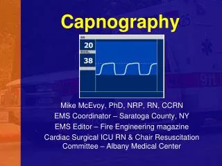
Definition of Capnography • A numerical value of the EtCO2 AND A waveform of the concentration of CO2 present in the airway • Respiratory rate detected from the actual airflow
Capnometer • A Capnometerprovides only a numerical measurement of carbon dioxide.
Capnogram • A Capnogram is a waveform display of carbon dioxide over time
Oxygenation and Ventilation What is the difference?
Oxygenation • Measured by pulse oximetry (SpO2) • Noninvasive measurement • Percentage of oxygen in red blood cells • Changes in ventilation take minutes to be detected • Affected by motion artifact, poor perfusion, etc.
Ventilation • Measured by the end-tidal CO2 • Partial pressure (mm Hg) or volume(% vol) of CO2 in the airway at the end of exhalation • Breath-to-breath measurement provides information within seconds • Not affected by motion artifact , poor perfusion, etc.
Oxygenation and Ventilation • Respiratory Cycle = separate physiologic processes: • Oxygenation • Ventilation
Comparing Capnography with Pulse Oximetry • Capnography • Carbon dioxide • Reflects ventilation • Hypoventilation / apnea detected immediately • Reflects change in ventilation within 10 seconds • Should be used with pulse oximetry • Pulse oximetry • Oxygen saturation • Reflects oxygenation • SpO2 changes lag when patient is hypoventilating or apneic • Reflects change in oxygenation within 5 minutes • Should be used with capnography
Physiology • Relationship between CO2 and RR • RR CO2 Hyperventilation • RR CO2 Hypoventilation • There is an inverse relationship between your respiratory rate and your CO2 level. • As you breath faster your RR goes up, your body is unable to hold onto CO2 and therefore blows it off faster so those levels go down. • As you breath more slowly, your RR goes down, your body is holding more CO2 due to the lack of breaths taken and the CO2 level goes up.
Elements of a Waveform End of exhalation Alveolar Gas Alveolar gas mixes with dead space Inspiration
The Normal CO2 Waveform • A – B Baseline • B – C Expiratory Upstroke • C – D Expiratory Plateau • D ETCO2 value • D – E Inspiration begins
ETT • A normal capnogram is the best evidence that the ETT is correctly positioned • With an esophageal tube little or no CO2 is present
Hypoventilation(increase in ETCO2) • Possible causes: • Decrease in respiratory rate • Decrease in tidal volume • Increase in metabolic rate • Rapid rise in body temperature (hypothermia)
Waveform: Regular Shape, PlateauAboveNormal • Indicates increase in ETCO2 • Hypoventilation • Respiratory depressant drugs • Increased metabolism • Fever, pain, shivering • Interventions • Adjust ventilation rate • Decrease respiratory depressant drug dosages • Assess pain management • Conserve body heat
Hyperventilation(decrease in ETCO2) • Possible causes: • Increase in respiratory rate • Increase in tidal volume • Decrease in metabolic rate • Fall in body temperature (hyperthermia)
Waveform: Regular Shape, Plateau Below Normal • Indicates CO2 deficiency • Hyperventilation • Decreased pulmonary perfusion • Hypothermia • Decreased metabolism • Interventions • Adjust ventilation rate • Evaluate for adequate sedation • Evaluate anxiety • Conserve body heat
Obstruction-Shark Fin • Possible causes: • Partially kinked or occluded artificial airway • Presence of foreign body in the airway • Obstruction in expiratory limb of the breathing circuit • Bronchospasm-Can indicate need for bronchodilators.
Sudden Loss of Waveform • Apnea • Airway Obstruction • Dislodged airway (esophageal) • Airway disconnection • Ventilator malfunction • Cardiac Arrest
Curare Cleft • Curare cleft is when a neuromuscular blockade wears off • The patient takes small breaths that causes the cleft • Management: consider neuromuscular blockade re-administration
TroubleshootingSudden increase in EtCO2 Malignant Hyperthermia Ventilation of previously unventilated lung Increase of blood pressure Release of tourniquet Bicarb causes a temporary <2 minute rise in ETCO2
TroubleshootingEtCO2 values drop to 0 Extubation/Movement into hypopharynx Ventilator disconnection or failure EtCO2 defect ETT kink
TroubleshootingSudden decrease EtCO2 (not to 0) Leak or obstruction in system Partial disconnect Partial airway obstruction (secretions) High-dose epi can cause a decrease
TroubleshootingContinual, exponential decrease in EtCO2 Pulmonary Embolism Cardiac Arrest Sudden hypotension/hypovolemia Severe hyperventilation
What does it really do for me? • Non-Intubated Applications • Bronchospasms: asthma, COPD, anaphlyaxis • Hypoventilation: drugs, stroke, CHF, post-Ictal • Shock and circulatory compromise • Hyperventilation Syndrome: biofeedback • Intubated Applications • Verification of ETT placement • ETT surveillance during transport • Control ventilations during CHI and increased ICP • CPR: compression efficacy, early signs of ROSC, survival predictor
MICROSTREAM CAPNOGRAPHY SOLUTIONS • Small pin holes deliver pillow of oxygen around both nose and mouth Nasal and Oral Sampling • CO2 sampling / O2 delivery for non-intubated patients • Uni-junction™ of sampling ports prevents dilution from nonbreathing source • Increased surface area provides greater sampling accuracy in the presence of low tidal volume
Smart CapnoLine™ Plus O2nasal cannula for CO2 measurement and O2 delivery • O2 delivery method reduces CO2 sampling dilution • –Solution for high flow O2 delivery (works effectively under oxygen delivery mask)
FilterLine® patient interfaces Prevents moisture from entering monitor Replaces water trap
Capnography in procedural sedation • Accurately monitors RR • Monitors adequate ventilation with non-intubated patients • Monitors potential risk of over-sedation resulting in • Hypoventilation more effectively than pulse oximetry • Early indicator of airway obstruction • Early warning of apnea • Adds an additional level of patient safety providing the caregiver with vital information to make accurate assessments and timely interventions for the patient