Chromosomes
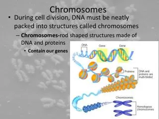
Chromosomes. Dr Pupak Derakhshandeh, PhD Ass Prof Medical Science of Tehran University. What are chromosomes?. Chromosomes are the structures that hold our genes Genes are the individual instructions that tell our bodies how to develop and function

Chromosomes
E N D
Presentation Transcript
Chromosomes Dr Pupak Derakhshandeh, PhD Ass Prof Medical Science of Tehran University
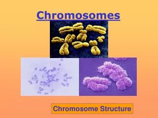
What are chromosomes? Chromosomes are the structures that hold our genes Genes are the individual instructions that tell our bodies how to develop and function They govern our physical and medical characteristics, such as hair color, blood type and susceptability to disease. Each chromosome has a p and q arm; p is the shorter arm and q is the longer arm. The arms are separated by a pinched region known as the centromere
How many chromosomes do humans have? • The typical number of chromosomes in a human cell is 46 - two pairs of 22 + XX/XY • Holding an estimated 30,000 to 35,000 genes. • One set of 23 chromosomes is inherited from the biological mother (from the egg), and the other set is inherited from the biological father (from the sperm).
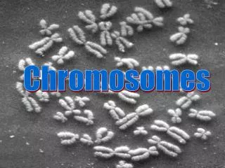
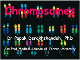
study of the chromosomes • with a microscope, then Stainning • The chromosomes look like strings with light and dark "bands" • A picture, or chromosome map, of all 46 chromosomes is called a karyotype • The karyotype : identify chromosome abnormalities: that are evident in either the structure or the number of chromosomes.
study of the chromosomes • The pairs have been numbered from 1 to 22, with the 23rd pair labeled "X" and "Y." • In addition, each chromosome arm is defined further by numbering the bands that appear after staining • The higher the number, the further that area is from the centromere.
Study of the chromosomes • The first 22 pairs of chromosomes are called "autosomes" • Final pair is called the "sex chromosomes." • The sex chromosomes an individual has determines that person's gender; females have two X chromosomes (XX), and males have an X and a Y chromosome (XY)
How Chromosome Abnormalities Happen? Meiosis Mitosis Maternal Age Environment
Meiosis • Chromosome abnormalities : • happen as a result of an error in cell division. • “Meiosis” : the cell division that the egg and sperm go through when they are developing. • Normally, meiosis causes a halving of chromosome material, so that each parent gives 23 chromosomes to a pregnancy
Chromosome abnormalities • Abnormality of chromosome number or structure: • Numerical Abnormalities • Structural Abnormalities
Numerical Abnormalities • When an individual is missing either a chromosome from a pair (monosomy) or has more than two chromosomes of a pair (trisomy). • An example: Down Syndrome, also known as Trisomy 21 (an individual with Down Syndrome has three copies of chromosome 21, rather than two). • Turner Syndrome is an example of monosomy the individual is born with only one sex chromosome, an X. • Kleinfelter Syndrome is an example of trisomy the individual is born with three sex chromosome, XXY.
Down Syndrome (Trisomy 21( Trisomy 2(
Down syndrom) Trisomy 21, 46) • critical region: • A region on the long (q) arm of chromosome 21 • Down syndrome causes mental retardation, a characteristic facial appearance, and multiple malformations • Associated with a major risk for heart malformationsa small but still significant risk of acute leukemia . • 3 copies of chromosome number 21
incidence of 1 in 660 and is by far the most common chromosomal abnormality • Slight flattening of the face • A low bridge of the nose (lower than the usually flat nasal bridge of the normal newborn) • An epicanthal fold (a fold of skin over top of the inner corner of the eye, which can also be seen less frequently in normal babies) • A ring of tiny harmlesswhite spots around the iris • mental retardation
Trisomy 18, 47 Ch. • incidence of about 1 in 3,000 • There is a 3:1 preponderance of females to males • Thirty percent of affected newborns die within the first month • 50% by two months • and 90% by one year. • severe mental retardation • microcephaly • overlapping fingers, and rocker bottom feet • Neurologically they are hypertonic • Other common malformations include congenital heart, kidney, .... abnormalities.
Trisomy 13 (XX/XY, 47 Ch) • has an incidence of 1 in 5,000 • Forty-four percent of affected newborns succumb in the first month of life • and 69% by six months • Only 18% of the babies born with trisomy 13 survive the first year • microcephaly • microophthalmia (small eyes) • cleft lip or cleft palate • polydactyly (extra fingers) • congenital heart defects • urogenital defects • brain malformations • severe mental retardation.
Turner Syndrome ( 45, X) 45, X
Turner syndrome • Only females • One X chromosome • Or has two X chromosomes but one is damaged • Short stature • Delayed growth of the skeleton • Sometimes heart abnormalities • Usually infertile due to ovarian failure • Diagnosis is by blood test (karyotype) • 1 out of every 2,500 female live births worldwide • Short neck with a webbed appearance
Kleinefelter XXY
Klinefelter syndrome (47, XXY) • In boys and men • 47 chromosomes with XXY sex chromosomes • XXY is one of the most common chromosomal abnormalities • 1 in 500 male births • the most common genetic cause of male infertility • Often : undiagnosed : variation in clinical presentation • Small testes , insufficient production of testosterone , and infertility
Klinefelter syndrome (47, XXY) • Breast enlargement, lack of facial and body hair, a rounded body type , to be overweight , and be taller than their fathers and brothers • Learning and/or behavioral problems • Testosterone replacement corrects the symptoms of androgen deficiency • Sex chromosome and autosomal chromosome abnormalities is higher in spermatozoa from patients with Klinefelter syndrome than in those from normal men.
Fragile X Syndrome • 1 in 3,600 males and 1 in 4,000 to 6,000 females with the full mutation worldwide • It is estimated that 1 in 250 females and 1 in 700 males are carriers of the premutation. • It is second only to Down Syndrome as a cause of mental retardation • Fragile X syndrome appears in children of all ethnic, racial and socio-economic backgrounds
Fragile X Syndrome • most common inherited form of familial mental retardation • (CGG)n trinucleotide expansion in the FMR1 gene leading to the typical Martin-Bell phenotype • Clinical features vary depending on age and seX • Expansion of a (CCG)n repeat in the FMR2 gene corresponds to the FRAXE fragile site which lies distal to FRAXA and is also associated with mental retardation, but it is less frequent and lacks a consistent phenotype
Chromosome abnormalities • Abnormality of chromosome number or structure: • Numerical Abnormalities • Structural Abnormalities
Structural Abnormalities • Deletions: A portion of the chromosome is missing or deleted (>5 Mb). • Paraderwilli Syndrome (Ch 15) • Angleman Syndrome (Ch 15) • Imprinting effect
DELETIONS • Deletion refers to the loss of a segment of a chromosome • This can be terminal (close to the end of the chromosome on the long arm or the short arm) • or it can be interstitial (within) • eg.DGS II
Structural Abnormalities • Duplications: A portion of the chromosome is duplicated, resulting in extra genetic material. • Oncogenes (c-onc, c-fos, c-myc)
DUPLICATIONS • refers to an extra chromosomal segment within the same homologous chromosome or an extra chromosomal segment on another nonhomologous chromosome. • Again, the clinical findings are highly variable depending upon the chromosomal segments involved. • Gene expantion: • in Huntington Disease/ Fragile X, ….
Structural Abnormalities • Translocations: When a portion of one chromosome is transferred to another chromosome.
There are two main types of translocations. • In a reciprocal translocation, segments from two different chromosomes have been exchanged. In a Robertsonian translocation, an entire chromosome has attached to another at the centromere.
TRANSLOCATIONS • Translocation involves two nonhomologous chromosomes (e.g., chromosome 2 and chromosome 6) • Following a break in each of the chromosomes, and subsequent reunion • a segment of chromosome 2 becomes attached to chromosome 6
Balanced reciprocal translocation Balanced reciprocal translocation Balanced reciprocal translocation