Download
1 / 33
330 likes | 474 Views
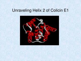
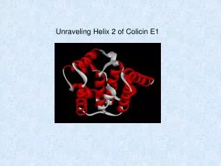
Unraveling Helix 2 of Colicin E1. Background. Zakharov et al., BBA. 2004. family of antimicrobial proteins secreted upon environmental stress (regulated by SOS system) three modes of action: 1. depolarization through ion channels 2. inhibition of protein and

E N D
Zakharov et al., BBA. 2004 family of antimicrobial proteins secreted upon environmental stress (regulated by SOS system) three modes of action: 1. depolarization through ion channels 2. inhibition of protein and peptidoglycan synthesis 3. degradation of nucleic acids 1994 – crystal structures of polypeptide fragments of Col E1 1997 – complete crystal structure of channel-forming P190 fragment
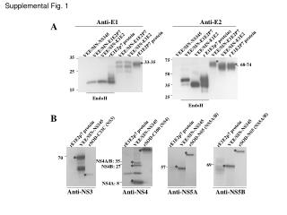
Cleavage sites: Zhang and Cramer, 1992 Palmer and Merrill, JBC, 1994 Parallax method of depth dependent fluorescence quenching of trp Y367 classed as moderately buried
Tory and Merrill, JBC, 1999 Fluorescence and FRET study red Δ λ emission max (W) blue reduced accessibility bimolecular quench constant lifetime Y367 immersed or interfacial
no REES exposed or embedded no REES buried moderate to large REES interfacial Tory and Merrill, BBA, 2002 Red-edge excitation shift analysis Y367 interfacial and sequestered from aqueous solvent
Musse et al., sometime soon! Helix one displayed an alpha-helical nature in both soluble and membrane-bound states. No elongation or blending of helices 1 and 2. These results support the toroidal pore model.
Zakharov and Cramer, BBA, 2002 Zahkarov et al., Biophys. J., 2004 Zakharov and Cramer, BBA, 2002 Model for colicin import Colicin membrane insertion
Shai, BBA, 1999 Zakhorov et al., BBA, 2004 Zakharov and Cramer, BBA, 2002 Models for membrane insertion
E365 D376 Q372 K379 K369 G380 S368 E373 A 375 K366 A371 K377 S378 M370 Y367 L374 hydrophobic polar acidic basic Helix 2 of Colicin E1
S378 G380 P190H
mBBr • well characterized • relatively small (about the size of a tryptophan) • uncharged, non-perturbing (structure or binding) • essentially non fluorescent until conjugated • fluorescence quenched by near-by W and, to a lesser extent, Y
Intrinsic Trp Fluorescence • indication of folded integrity of mutant protein compared with WT • using 295 nm excitation avoids fluorescence from the 9 Y residues • the three trp residues are in rigid environments and exhibit limited • flexibility
protein TX-100 SPQ in vitro Channel Assay • test of the pore-forming ability of the mutant protein
Bimane Fluorescence • reports on accessibility of bimane probe which relates to “location” within • the tertiary structure of the protein standard apparent polarity scale curve bimane-N-acetyl-Cys dioxane-water solvent system Musse and Merrill
Surface Area Solvent Accessibility GETAREA 1.1 Solvent Accessible Surface Areas, Atomic Solvation Energies, and Their Gradients for Macromolecules Sealy Center for Structural Biology, University of Texas Medical Branch, Galveston, TX 77555 Area Per residue Job identifier: get_a_13306 Probe radius : 1.400 Residue Total Apolar Backbone Sidechain Ratio(%) In/Out ILE 345 121.48 109.19 13.62 107.86 73.2 o LYS 346 151.45 27.52 15.63 135.82 82.6 o ASP 347 102.95 34.60 0.13 102.82 91.0 o ALA 348 6.96 6.68 3.62 3.34 5.1 i VAL 349 74.04 74.04 0.00 74.04 60.5 o ASP 350 89.67 30.09 8.52 81.15 71.8 o ALA 351 36.79 35.19 7.92 28.87 44.5 THR 352 0.04 0.04 0.00 0.04 0.0 i VAL 353 89.36 89.33 0.13 89.23 73.0 o SER 354 68.44 41.93 7.49 60.95 78.7 o
Anisotropy • predicts local environment within tertiary structure by measuring rotational • property of bimane moiety on cysteine residue • low value = free moving • high value = restricted movement
>30% 10-25% <5%
Quantum Yield • = number of photons emitted/the number of photons absorbed • = fraction of fluorophore that decays through emission • low value = accessible • high value = not accessible
>30% 10-25% <5%
depth study • quenching with KI (shallow) and 10-DN (deep) • quenching is insensitive to variation in lipid content • 10-DN retains “free energy” over range of bilayer depth • could do study with each quencher alone, but ratio between shallow and • deep increases sensitivity and eliminates non-depth related quenching • effects (excited state lifetime of bimane) • ratio is linearly dependent on depth of trp residue Dual Quenching Assay Erwin London
>0.4 0.11-0.4 <0.1 low ratio value = accessible high ratio value = buried
Summary Bimane lambda emission max (membrane-bound vs soluble): Blue shifted (455-460 nm): A371, S378 Intermediate (461-470 nm): Y367, K369, M370, L374, A375, K377 Red shifted (>471 nm): S368, Q372, E373, D376, K379, G380 Soluble anisotropy: Accessible (0.095-0.135): K369, M370, A371, E373, D376, K377, K379, G380 Inaccessible (0.157-0.192): Y367, S368, Q372, L374, A375, S378 These residues are considered buried, but mBBr may be mobile within “pocket” of protein structure. These residues are considered exposed, but probe may be immobilized by tertiary structure contacts. Membrane bound anisotropy: Little change (<5%): Y367, S368, Q372, A375 Moderate change (10-25%): K369, E373, L374, D376, S378, K379, G380 Significant change (>30%): M370, A371, K377 Quantum Yield: Little change (<5%): K369, M370, E373, L374, D376, K379 Moderate change (10-25%): Y367, Q372, K377, G380 Significant change (>30%): S368, A371, A375, S378 S368 and A371 had rather low soluble QF values (<0.18, the observed value for Cys-bimane standard) which may have been caused by quenching of the bimane signal by Y367. Dual Quenching Analysis: Accessible (<0.10): Y367, S368, K369, Q372, D376, K377 In-between (0.11-0.4): E373, L374, S378, K379, G380 Buried (>0.4): M370, A371, A375