Download
1 / 63
640 likes | 824 Views
The Basics of Mitosis & Meiosis. GUTS Lecture James P. Evans MD, Ph.D Fall 2008. Objectives. Understand the process of somatic cell division: mitosis -> daughter cells germ cell division: meiosis -> gametes The consequences of these processes and resulting errors. What is Mitosis?.

E N D
The Basics of Mitosis & Meiosis GUTS Lecture James P. Evans MD, Ph.D Fall 2008
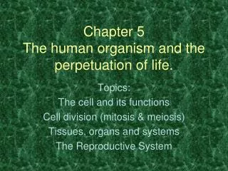
Objectives Understand the process of • somatic cell division: mitosis -> daughter cells • germ cell division: meiosis -> gametes • The consequences of these processes and resulting errors
What is Mitosis? • Somatic cell division consisting of nuclear division & cytoplasmic division (cytokinesis) • Single cell divides to produce two identical daughter cells
“You cannot step twice into the same river”…Heraclitus of Ephesus, c.535 BCE-475 BCE) • You literally are not the same person you were in the past • Cell division is required for • Growth & Development • Human zygote = 1 cell • Mature adult = 1 X 10¹⁴ cells • We wear out • Cell division is a primary manner in which we deal with the harmful effects of wear and damaging forces in our environment • Stem cells form a pool of self-renewing cells as “replacement parts”
What do all cells require to survive? • A complete set of genetic instructions in order to • produce required molecules • direct life processes • We need an orderly process to duplicate and distribute chromosomes through successive cell divisions
Mitosis • Fundamental process by which cells and DNA replicate • Diploid (2n) cell > two diploid daughter cells • Maintains chromosome # • One cell with 46 chromosomes > two cells each with 46 chromosomes • Maintains DNA content • One cell with 2n DNA > two cells each with 2n DNA
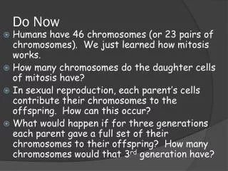
Mitosis • Diploid organisms receive one chromosome from their female parent (maternal chromosome) and one chromosome from male parent (paternal chromosome) • A “matched” pair of maternal and paternal chromosomes are called homologues • Humans - 23 pairs of homologous chromosomes • 22 pairs of autosomes and 1 pair of sex chromosomes
Cell Cycle cell grows in size organelles replicated Interphase = G1, S, G2 (non dividing) G1 10-12 hrs S replication of DNA, synthesis of proteins associated with DNA Mitotic Cell Division 6-8 hrs G2 synthesis of proteins associated with mitosis 2-4 hrs
arm arm centromere duplicated chromosome Unreplicated Chromosome • S phase • Chromosomes (DNA) are replicated 2 sister chromatids – Attached at their centromeres
Duplicated chromosome 2 sister chromatids 2 daughter chromosomes
The Phases of Mitosis 4 phases: 1st – Prophase 2nd – Metaphase 3rd – Anaphase 4th – Telophase/Cytokinesis
chromatin nucleus centrioles Prophase nucleolus • Centrioles duplicated to form two pairs • Chromosomes begin to condense
condensing chromosomes Prophase • Nuclear envelope • begins to break down • Spindle fibers form (specialized microtubules) • Radiate out from centrioles • forming the “aster” • One pair of centrioles migrates to • one pole of cell, the other pair • migrates to opposite pole of cell
The Mitotic Spindle • Spindle fibers are specialized microtubules • Spindle fibers radiate out from centrioles forming the “aster” • Centrioles occur in pairs, and are duplicated during interphase
Prophase The Spindle Captures Chromosomes • When spindle fibers are fully formed nuclear envelope disintegrates & nucleolus disappears • Spindle fibers attach to chromosomes at the kinetochore, a structure located at the centromere Duplicated chromosome (2 sister chromatids)
Prophase • Function of spindle fibers is to organize division of sister chromatids into daughter cells • Other spindle fibers do NOT attach to chromosomes, but retain free ends that overlap at cell’s equator > “free spindle fibers”
Metaphase Chromosomes align along equator of the cell, with one kinetochore facing each pole centrioles chromosomes spindle fibers
Anaphase • Sister chromatids separate • Spindle fibers attached to kinetochores shorten and pull chromatids poleward • free spindle fibers lengthen and push poles of cell apart
Telophase Chromosomes decondense spindle fibers disintegrate pinching of cell membrane at equator nuclear envelope reforms nucleolus reappears
microfilaments attach to plasma membrane to form a ring around equator of cell cytokinesis ring contracts, like a drawstring, dividing the cytoplasm Daughter Cells
Mitotic Errors • Mitotic nondisjunction • Failure of sister chromatids to separate • results in chromosome imbalance
Chromosome Imbalance Mitotic nondisjunction: failure of sister chromatids to disjoin
45, XX, -21 47, XX, +21 Mitotic Nondisjunction After Fertilization -fertilization Monosomic 46, XX Trisomic Mitotic nondisjunction in one cell 46, XX 46, XX Daughter cells do not have identical genetic content = mosaic 46, XX 2-cell embryo
Nature Genetics 3, 146 - 150 (1993) Mitotic errors in somatic cells cause trisomy 21 in about 4.5% of cases and are not associated with advanced maternal age Stylianos E. Antonarakis, Dimitrios Avramopoulos, Jean-Louis Blouin, C. Conover Talbot Jr & Albert A. Schinzel
Vol. 94, No. 1-2, 2001 Frequent mitotic errors in tumor cells of genetically micro-heterogeneous glioblastomas S. Loepera, B.F.M. Romeikeb, N. Heckmanna, V. Junga, W. Henna, W. Feidenb, K.D. Zanga, S. Urbschata Glioblastoma multiforme (GBM) is characterized by intratumoral heterogeneity as to both histomorphology and genetic changes, displaying a wide variety of numerical chromosome aberrations the most common of which are monosomy 10 and trisomy 7.
Mitosis Orderly duplication & distribution of chromosomes through successive somatic cell divisions
Problem The number of chromosomes is constant from cell to cell …So how does the union of sperm and egg produce the same number of chromosomes found in cells of the parents?
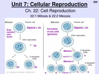
Mitosis One diploid (2n) cell --> two diploid daughter cells Maintains chromosome # 46 --> 46 Maintains DNA content 2n --> 2n Meiosis One diploid (2n) --> Four haploid (1n) cells Reduces chromosome # from 46 --> 23 Reduces the DNA content from 2n --> 1n
X Meiosis Diploid germ cell Chromosome #: 46 DNA content: 2n Homologous chromosomes 2n Chromosome #: 46 DNA content: 4n 4n Reduction division Humans have 23 pairs of homologous chromosomes 22 pairs of autosomes 1 pair of sex chromosomes Chromosome #: 23 DNA content: 2n 2n Mitotic division Chromosome #: 23 DNA content: 1n Haploid gametes M P 1n chromosome 2 Fig. 2.6, page 9
2n 4n Reduction division Mitotic division 2n Haploid gametes 1n Meiosis Diploid germ cell Chromosome #: 46 DNA content: 2n Homologous chromosomes Chromosome #: 46 DNA content: 4n Chromosome #: 23 DNA content: 2n Chromosome #: 23 DNA content: 1n Fig. 2.6, page 9
2n 4n Reduction division Mitotic division 2n Haploid gametes 1n Meiosis Diploid germ cell Chromosome #: 46 DNA content: 2n Homologous chromosomes Chromosome #: 46 DNA content: 4n Chromosome #: 23 DNA content: 2n Chromosome #: 23 DNA content: 1n Genetic variation is generated by random assortment of homologous chromosomes during the reduction division Fig. 2.6, page 9
2n 4n Reduction division Mitotic division 2n Haploid gametes 1n Meiosis Diploid germ cell Chromosome #: 46 DNA content: 2n Homologous chromosomes Chromosome #: 46 DNA content: 4n Chromosome #: 23 DNA content: 2n Chromosome #: 23 DNA content: 1n
The Four Phases of Meiosis I • Cell division that reduces the chromosome number by one-half • four phases: a. prophase I b. metaphase I c. anaphase I d. telophase I
Homologous chromosomes sister chromatids sister chromatids Tetrad Prophase I - Synapsis maternal paternal
Crossing Over • Crossing over occurs between nonsister chromatids of homologous chromosomes at the chiasmata (point of crossing over) • Segments of nonsister chromatids break and reattach to the other chromatid
Tetrad Crossing Over Generates Genetic Variation nonsister chromatids P M M P M/P P/M variation chiasmata: site of crossing over
Consequences of Meiotic Recombination (Crossing Over) • exchange of genetic material between maternal & paternal homologous chromosomes • Generates genetic variation • Ensures normal chromosome disjunction
spindle fiber centrioles aster fibers Prophase I
Chromosome 2 P M M P P M OR Chromosome 3 metaphase plate metaphase plate Metaphase I • Tetrads align on the metaphase plate. Chromosome 2 M P Chromosome 3
Independent Assortment Generates Genetic Diversity • Orientation of homologous pair to poles is random • 223 = > 8 million combinations • Combining with crossing over results in > 6 x 10²⁰ unique possibilities for a single germ cell
Anaphase I • Homologous chromosomes • separate and move towards the poles. • Sister chromatids remain attached at their centromeres.
Telophase I • Cytokinesis occurs and two daughter cells are formed • haploid chromosome # (23) • 2n DNA • Completion of Reduction Division
Meiosis II • No interphase II • or very short - no more DNA replication • Meiosis II is similar to mitosis
Prophase II • same as prophase in mitosis
metaphase plate metaphase plate Metaphase II • same as metaphase in mitosis
Anaphase II • same as anaphase in mitosis • sister chromatids separate
Summary of Meiosis I and II • Meiosis I separates homologous chromosomes • Resulting in 2 daughter cells • Haploid chromosome content (#23) • Diploid DNA content (2n) • Meiosis II separates sister chromatids • Resulting ultimately in 4 daughter cells • Haploid chromosome content (# 23) • Haploid DNA content (1n)
Meiosis I Meiosis I Meiosis II Meiosis II Four haploid gametes One haploid gamete Meiosis Differs Between Males and Females Spermatogenesis Oogenesis polar body