Download
1 / 11
110 likes | 128 Views
Test your structure-spectrum correlation skills with NMR problems for amino acids. Learn to assign signals, identify isomers, and differentiate amino acids based on NMR spectra. Understand PPM units and carbon signal characteristics.

E N D
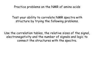
Practice problems on the NMR of amino acids Test your ability to correlate NMR spectra with structure by trying the following problems. Use the correlation tables, the relative sizes of the signal, electronegativity and the number of signals and logic to connect the structures with the spectra.
NMR units are ppm (parts per million). We discuss where this ppm unit comes from in class. These are the signals of protons (1H NMR). We think of the position (in ppm) of the NMR signals in the same way we viewed the frequency of IR signals. The 1H NMR signals are characteristic of certain types of protons in molecules
These are the position of the signals of carbon atoms (13C) in ppm. We think of the position of a NMR signal in ppm in the same way we viewed the frequency of IR signals. The 13C NMR signals are characteristic of certain types of carbon atoms in molecules.
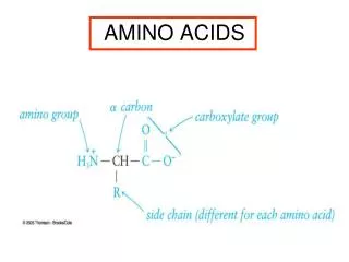
O H C H 3 H v a l i n e H O C H 3 N H 2 12 10 8 6 4 2 0 PPM The 1H NMR of the amino acid valine is given below. Assign the protons of valine to the signals (a, b, c, d and e) in the NMR spectrum. a b c d d The answer is at the end of the ppt
180 160 140 120 100 80 60 40 20 0 PPM 180 160 140 120 100 80 60 40 20 0 PPM Leucine and isoleucine are isomers. Two 13C NMR spectra A and B are shown below. One is leucine the other is isoleucine. Which is which? A B
180 160 140 120 100 80 60 40 20 0 PPM 180 160 140 120 100 80 60 40 20 0 PPM The 13C NMR spectrum of valine and leucine are shown. Which is valine and which is leucine? valine signal signal leucine
180 160 140 120 100 80 60 40 20 0 PPM 180 160 140 120 100 80 60 40 20 0 PPM The 13C NMR spectrum of phenylalanine and tyrosine (both possess aromatic rings) are shown. Which is phenylalanine and which is tyrosine? phenylalanine Two signals Very close together tyrosine
O H C H 3 H v a l i n e H O C H 3 N H 2 12 10 8 6 4 2 0 PPM The 1H NMR of the amino acid valine is given below. Assign the protons of valine to the signals in the NMR spectrum. a a e d c a b b d c e
180 160 140 120 100 80 60 40 20 0 PPM 180 160 140 120 100 80 60 40 20 0 PPM Leucine and isoleucine are isomers. Two 13C NMR spectra A and B are shown below. One is leucine the other is isoleucine. Which is which? Two equivalent carbon atoms (the two CH3 groups) A B All the carbon atoms are unique
180 160 140 120 100 80 60 40 20 0 PPM 180 160 140 120 100 80 60 40 20 0 PPM The 13C NMR spectrum of valine and leucine are shown. Which is valine and which is leucine? An essential difference between valine and leucine is that leucine has one more carbon atom. In the bottom spectrum the CH2 carbon atom of leucine is just to the left of the CH3 carbon atom. valine signal signal leucine
tyrosine 180 160 140 120 100 80 60 40 20 0 PPM Two signals Very close together phenylalanine 180 160 140 120 100 80 60 40 20 0 PPM The 13C NMR spectrum of phenylalanine and tyrosine (both possess aromatic rings) are shown. Which is phenylalanine and which is tyrosine? The most significant difference between the two structures is the OH on the benzene ring of the tyrosine. This substituent will cause the carbons of the benzene ring of tyrosine to be much more different chemically than those of phenylalanine. This difference will also be reflected in the differences in the separation of the carbon signal in the NMR