Download
1 / 36
360 likes | 525 Views
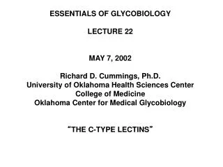
ESSENTIALS OF GLYCOBIOLOGY LECTURE 24 MAY 9, 2002 Richard D. Cummings, Ph.D. University of Oklahoma Health Sciences Center College of Medicine Oklahoma Center for Medical Glycobiology “ THE PLANT LECTINS ”. “ THE PLANT LECTINS ”. Definition of a Lectin -

E N D
ESSENTIALS OF GLYCOBIOLOGY LECTURE 24 MAY 9, 2002 Richard D. Cummings, Ph.D. University of Oklahoma Health Sciences Center College of Medicine Oklahoma Center for Medical Glycobiology “THE PLANT LECTINS”
“THE PLANT LECTINS” Definition of a Lectin - “A protein (other than an anti-carbohydrate antibody) that specifically recognizes and binds to glycans without catalyzing a modification of the glycan.” The first lectins identified were derived from plants, specifically leguminous seeds. Until recently, it was thought that a lectin must be multivalent and soluble. But some monovalent, monomeric lectins, and many membrane-bound lectins, are now known.
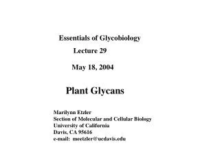
History of Plant Lectins Date Investigators Discovery • 1888 H. Stillmark Ricinus communis plant extract • has hemagglutinating properties • 1890 P. Ehrlich Lectins used as antigens in early • Immunological studies • 1908 K. Lansteiner & Different hemagglutinating • H. Raubitsheck properties in various plant seeds • 1919 J. Sumner Crystallization of Con A • 1936 J. Sumner Lectins bind sugar - Con A • precipitates glycogen
Datura stramonium (jimsonweed) Ricinus communis (castor bean) Lycopersicum esculentum (tomato) Lens culinaris (lentil)
History of Plant Lectins Date Investigators Discovery • 1940 W. Boyd, Lectins specific for some human R. Reguera & blood group antigens • K.O. Renkonen • 1952 W. Watkins & Use of lectins and glycosidases to W. Morgan prove that blood group antigens • are sugars and to deduce the • structures of the antigens • 1954 W. Boyd & The name lectin is proposed to E. Shyleigh replace hemagglutinin
History of Plant Lectins Date Investigators Discovery • 1960 P.C. Nowell Red kidney bean lectin P. • & J.C. Aub vulgaris mitogenic for resting • lymphocytes • 1960’s M. Burger Lectins preferentially • 1970’s G. Nicolson agglutinate some animal tumor • cells • 1980’s Kornfeld(s) Use of immobilized lectins • Osawa to analyze animal • Kobata glycoconjugates • Cummings • 1980’s D. Kabelitz Discovery that plant lectins • 1990’s D.J. Gee induce apoptosis • K. Schweizer
SOME FAMILIES OF LECTINS DISTINGUISHED BY 3º STRUCTURE • Lectin group Structure of CRD Length • Calnexin Unknown ? • L-type b-sandwich ~230 (Legume lectin-like) • P-type Unique b-rich structure ~130 (Phosphomannose) • M-type Unique a-helical ~500 (mannosidase-related • C-type Unique mixed /ß structure ~115 (Ca2+-dependent) • Galectins b-sandwich ~125 • I-type Immunoglobulin superfamily ~120 • R-type b-trefoil (plants and animals) ~125 (Ricin related)
Uses of Plant Lectins • Agglutination of cells and blood typing • Cell separation and analysis • Bacterial typing • Identification and selection of mutated cells with • altered glycosylation • Toxic conjugates for tumor cell killing • Cytochemical characterization/staining • of cells and tissues • Mitogenesis of cells • Mapping neuronal pathways • Purification and characterization of glycoconjugates • Assays of glycosyltransferases and glycosidases • Defining glycosylation status of target glycoconjugates a
Primary Structural Motifs in Leguminous (L-type) Plant Lectins N-TERMINI 1 Lima bean lectin Phaseolus limensis A-E-L-F-F-N-F-Q-T-F-N-A-A-N- 1 SBA - Soybean agglutinin (Glycine max) A-E-T-V-S-F-S-W-N-K-F-V-P-K-Q- FYWKR EDQ LIV LIV Conserved Motif In C-terminal Domain FL ST Red = invariant residues - -x- - -V-x- -G- - - Lima bean lectin -V-L-D-D-W-V-S-V-G-F-S-A- SBA -S-L-P-E-W-V-R-I-G-F-S-A- STAG FLI EQV LIV METAL BINDING SITES ST - - -V- - -D- - Lima bean lectin -L-T-V-A-V-E-F-D-T-C-H-N- -Q-V-V-A-V-E-F-D-T-F-R-N- SBA
Classifications of Some Plant Lectins Binding Sites per Subunit Monosaccharide Specificity Subunit MW (kDa) Subunits Class Diverse 25-30 2 or 4 1 Legumes Grains Primarily Amino Sugars (GlcNAc/NeuAc) ~18 2 2 -S-S- Bonds Glycosylation Metals Class Variable No Mn2+, Ca2+ Legumes Grains Variable Yes No a
Similarities in Protein Folding Between Galectins and Legume L-type Lectins Con A Dimer Bovine Galectin-1 Dimer
Protein Folding in L-type Lectins Crystal structure of artocarpin lectin from the jack fruit (Artocarpus integrifolia) (left - monomer; right - tetramer)
Structure of L-type Tetrameric ConA at 2.35 Å. The trimannoside ligand is indicated in space-filling mode and the coordinated Ca2+ and Mn2+ are shown as the large green balls and small pink balls, respectively. The crystal structure was originally reported as a complex of ConA and a trimannoside ligand by Naismith and Field (Naismith J.H. and Field R.A. 1996. Structural basis of trimannoside recognition by concanavalin A. J. Biol. Chem. 271: 972–976).
Çrystal Structure of the L-type Dioclea guianensis Seed Lectin Ribbon representation showing the overall structure of Dioclea guianensis Seed Lectin tetramer and the relative location of the metal ions in the four subunits. The Mn2+ (green) and Ca2+ (yellow) of the canonical (S1 and S2) metal-binding site are shown as spheres. The secondary sub-sites for the Ca2+ /Cd2+ (S3) and Mn2+ (S5) are depicted as purple and blue spheres, respectively. (Ref: Wah et al, (2001) J. Mol. Biol. Vol. 310
Crystal Structure of “Grain-type” Wheat Germ Agglutinin (Isolectin 2) Dimer in Complex With N-Acetylneuraminyllactose Sugar binding site sialyllactose Wright CS (1990) 2.2 A resolution structure analysis of two refined N-acetylneuraminyl-lactose--wheat germ agglutinin isolectin complexes J Mol Biol 215, 635-651
Because of their multivalency and oligomeric structures, many plant lectin can cross-linking can precipitate glycoproteins and agglutinate cells
R R R R R R R R R R R C C C C C C C C C R-type CRD R-type CRD Hydrolase Domain GalNAcT Domain Ricin-type R-type Lectins - b-trefoil proteins Drosophila Lectins Bacterial Lectins Bacterial Hydrolases Ricin/Plant Toxins GalNAc Transferases Mannose Receptor Family C-type CRD Fibronectin domain TM domain
Structures of R-type Lectins Comparisons between Cys-MR (R-type domain in the mannose receptor) and other b-trefoil proteins - Cys-MR, a portion of the ricin B chain (residues 1–136 with N-linked carbohydrates omitted; and human aFGF (from Liu Y et al. (2000) J. Exp. Med., 191:1105-16)
Crystallographic structures of ricin (A) and Shiga toxin (B) The plant toxin ricin consists of two disulfide-linked polypeptides with different functions. The A-chain enters the cytosol and inactivates the ribosomes enzymatically (the A chain of ricin has RNA N-glycosidase activity to cleave a specific adenine base from ribosomal RNA), whereas the B-chain has lectin properties and binds to carbohydrates at the cell surface. (The structures have been obtained from the PDB protein data bank (ricin: 1DMO; Shiga toxin:2AA1), and are based on work published by Rutenber et al. (1991) and Fraser et al. (1994).)
Crystallographic structures of ricin (A) and Shiga toxin (B) This binding is a requirement for translocation of the A-chain to the cytosol. The bound toxin is endocytosed and transported retrograde through the Golgi apparatus to the endoplasmic reticulum where it appears to be translocated to the cytosol by the sec61p complex. (ref: Olsnes S, Kozlov JV. (2001) Ricin. Toxicon 39(11):1723-8). The cytosolic target of ricin and Shiga toxin is the 28S RNA of the 60S ribosomal subunit (Endo et al., 1987). Reduction of the disulfide bond connecting the A- and B-moieties of ricin is required for optimal enzymatic activity.
Lectin Biosynthesis • During biosynthesis, some of the leguminous lectins are proteolytically cleaved to generate a b-chain, corresponding to the amino terminus, and an a-chain, corresponding to the carboxyl terminus. • For example, jacalin lectin, from the jackfruit Artocarpus heterophyllus, is a tetrameric two-chain lectin (65 kD) (molecular mass 65 kD) with an a-chain of 133 amino acid residues and a b-chain of 20-21 amino acid residues. • An exceptional situation occurs with the well-known lectin Con A from jack beans (Canavalia ensiformis). • Con A is generated as a glycoprotein precursor, but it is proteolytically processed; the propeptide with the N-glycan is removed; the two chains are transposed and rejoined with the formation of a new peptide bond to generate the intact protein. • Thus, with regard to other lectins, the mature amino terminus of ConA corresponds to an a-chain and the carboxyl terminus corresponds to a b-chain. • In sequence alignments with other lectins, ConA exhibits what is called “circular” homology.
Biological Functions of Plant Lectins • Seed storage proteins • Aid in maintaining seed dormancy • Defense against fungal, viral, and bacterial pathogens • Defense against animal predators • Symbiosis in lugumes • Transport of carbohydrates • Mitogenic stimulation of embryonic plant cells • Elongation of cell walls • Recognition of pollen a
Plant Lectin Function in Nitrogen Fixation/Rhizobial Infection The roots of the legume Dolichos biflorus contain a lectin/nucleotide phosphohydrolase (Db-LNP) that binds to the Nod factor signals produced by Nod genes in rhizobia that nodulate this plant. Db-LNP is differentially distributed along the surface of the root axis in a pattern that correlates with the zone of nodulation of the root. Db-LNP is present on the surface of young and emerging root hairs and redistributes to the tips of the root hairs in response to treatment of the roots with a rhizobial symbiont or with a carbohydrate ligand. (Ref: Kalsi G, Etzler ME. (2000). Localization of a lipo-chitin oligosaccharides (LCOs), or Nod factors and Nod factor-binding protein in legume roots and factors influencing its distribution and expression. Plant Physiol 124(3):1039-48). Nod C encodes a GlcNAcT to synthesizes the chitin glycan; Nod B catalyzes the de-N-acetylation; Nod A catalyzes N-fatty acylation
OR HO OH OH OH HO H3C RO HO NHFatty Acid NHFatty Acid OH HO OH NAc NAc H3C OH OH OH HO NHHFatty Acid Plant Lectin Function in Nitrogen Fixation/Rhizobial Infection B F HO C E G A D Structure of lipo-chitin oligosaccharides in the pooled HPLC fractions 7 and 8 of Mesorhizobium loti strain NZP2213. Monosaccharide residues are labeled A-G. R1, predominantly C20:1 and C18:0, with other minor fatty acids; R2, carbamoyl NH2CO-; R3, acetyl or H. Olsthoorn et al, (1998) Biochemistry 37(25):9024-32
Some Uses of Plant Lectins • Agglutination of cells and blood typing • Cell separation and analysis • Bacterial typing • Identification and selection of mutated cells with altered glycosylation • Toxic conjugates for tumor cell killing • Cytochemical characterization/staining of cells and tissues • Mitogenesis of cells • Mapping neuronal pathways • Purification and characterization of glycoconjugates • Assays of glycosyltransferases and glycosidases • Defining glycosylation status of target glycoconjugates a
Example of a Catalog Listing (Vector Labs) Lectin Products Example - Aleuria Aurantia Lectin (AAL) • Agarose bound* Aleuria Aurantia Lectin (AAL) • Alkaline Phosphatase conjugated Aleuria Aurantia Lectin (AAL) • Biotinylated Aleuria Aurantia Lectin (AAL) • Unconjugated Aleuria Aurantia Lectin (AAL) • VECTREX AAL • VECTREX AAL Binding and Elution Kit
Serial Lectin Affinity Chromatography for Fractionation and Purification of Complex Carbohydrates Con A Quantity of Glycan Fraction Number LCA LCA SNA SNA L-PHA L-PHA Further Purification on Other Lectins, HPLC, etc.
Lectin Recognition of Glycans Mannose-Binding in N-Glycans [Hapten: 0.5 M a-Methyl-Man] [Hapten: 0.1 M a-Methyl-Man] [Hapten: 0.1 M a-Methyl-Glc]
a a a Lectin Recognition of Glycans Galactose-Binding in Complex-type N-glycans Bound By 1,4 1,2 b b Datura stramonium Gal GlcNAc Man 1,4 Man-GlcNAc-GlcNAc-Asn agglutinin (DSA) (weakly) b 1,2 b Gal GlcNAc Man Hapten: 10 mg/ml Chitotriose 1,4 1,4 b b Gal GlcNAc 1,4 b Gal GlcNAc 1,6 b b1,4 1,2 b Phaseolus vulgaris Gal GlcNAc Man Man-GlcNAc-GlcNAc-Asn leukoagglutinin (L4-PHA) 1,2 b1,4 b Gal GlcNAc Man Hapten: 0.4 M GalNAc GlcNAc 1,4 Phaseolus vulgaris b 1,2 b Gal GlcNAc Man 1,4 b erythroagglutinin (E4-PHA) Man-GlcNAc-GlcNAc-Asn 1,4 b 1,2 b Gal GlcNAc Man Hapten: 0.4 M GalNAc
Lectin Recognition of Glycans Galactose-Binding in Complex-type N- and O-glycans, and Glycosphingolipids Erythrina cristagalli lectin (specific for Galb4GlcNAc-R) Ricinus communis agglutinin (RCA-I) (binds better to Galb4GlcNAc-R than To Galb3GlcNAc-R ) Hapten for both: 0.1 M lactose Hapten: 10 mM raffinose Hapten: 50 mM GalNAc
Lectin Recognition of Glycans Fucose-Binding in Complex-type N- and O-glycans, and Glycosphingolipids Hapten: 0.2 M Fuc Hapten: 0.2 M Fuc Hapten: 0.2 M a-methyl-Man Hapten: 10 mM Fucose
Lectin Recognition of Glycans N-Acetylglucosamine-Binding in Complex-type N- and O-glycans, and Glycosphingolipids [Hapten:10 mg/ml Chitotriose] [Hapten: 0.1 M GlcNAc]
Lectin Recognition of Glycans Sialic acid-Binding in Complex-type N- and O-glycans, and Glycosphingolipids [Hapten: 50 mM Lactose] [Hapten: 50 mM Lactose]
Lectin Recognition of Glycans Galactose- and N-acetylgalactosamine-Binding In O-glycans [Hapten for all: 0.1 M GalNAc [Hapten for all: 50 mM a-Methyl-GalNAc]
Serial Lectin Affinity Chromatography for Fractionation and Purification of Complex Carbohydrates Con A Quantity of Glycan Fraction Number LCA LCA SNA SNA L-PHA L-PHA Further Purification on Other Lectins, HPLC, etc.
Biotin- Biotin- Biotin- Add Alk.Phos.- Streptavidin- Alk.Phos.- Streptavidin- Use of a lectin to assay a sialyltransferase in an ELISA-type Method Galb1-4GlcNAc-R- CMP-NeuAc a2-3-sialyltransferase Step 1 CMP NeuAca2-3Galb1-4GlcNAc-R- Add Biotinylated-MAL Step 2 NeuAca2-3Galb1-4GlcNAc-R- COLOR Step 3 NeuAca2-3Galb1-4GlcNAc-R-