방광 종양 (Bladder Tumor)
방광 종양 (Bladder Tumor). 동국대학교 비뇨기과 이 경 섭. 빈도. 우리 나라 비뇨기 암 중 가장 흔하다 . 인구 10 만 명 당 남자 7.76 명 , 여자 1.19 명 85%: localized bladder cancers 15%: regional LN or distant metastasis M : F = 2.7 : 1 white > black. Pathogenesis(1).


방광 종양 (Bladder Tumor)
E N D
Presentation Transcript
방광 종양(Bladder Tumor) 동국대학교 비뇨기과 이 경 섭
빈도 • 우리 나라 비뇨기 암 중 가장 흔하다. • 인구 10만 명 당 남자 7.76명, 여자 1.19명 • 85%: localized bladder cancers • 15%: regional LN or distant metastasis • M : F = 2.7 : 1 • white > black
Pathogenesis(1) • Multistep phenomenon of neoplastic change of urothelium - Initiator or its metabolites normal cell DNA → malignant cell 정상 genetic transformation의 변화 (DNA → RNA) → abnormal protein - Promoters: not carcinogenic proliferate already transformed cell
Pathogenesis(2) • Bladder cancer initiator or promoters 1) cigarette smoking - 50%(male), 31% (female) - α & β-naphthylamine in urine of smoker 2) occupational exposure - dye, rubber, petroleum, leather, printing - benzidine, β-naphthylamine, 4-aminobiphenyl 3) cytoxan 4> artificial sweetners 5) Schistosomiasis, instrumentation, calculi
Pathogenesis(3) • Activation of oncogenes c-Ha-ras, c-myc, c-ERB-2, Rb-1 • Tumor suppressor gene p53
Staging Jewett-Strong- Finding Marshall TNM병기 No tumor in the specimen 0 T0 Carcinoma in situ 0 Tis Noninvasivepapillary tumor 0 Ta Submucosal invasion A T1 Superficial muscle invasion B1 T2 Deep muscle invasion B2 T3a Invasion of perivesical fat C T3b Invasion of contiguous organ D1 T4 Regional lymph node metastases D2 (N1-3) Juxtaregional lymph node metastases D2 - Distant metastases D2 M1
Histopathology • Papilloma (Ta) - Papillary tumor with fine fibrovascular stalk - 2% of all TCC • Transitional cell Ca.(TCC) - 90% - papillary, exophytic lesion : superficial sessile: invasive
WHO grading (by Mostofi) Grade Ⅰ: 고분화형,작은 유두상 종양, 가는 줄기,다발성 경향, 경과가 좋고 예후가 양호 Grade Ⅱ: 중간분화형, 낮은 분화도, 핵 분열상,줄기가 있고 유두상의 종양이 대부분. grade I 종양에 비해 큰 경향이 있고 점막하 침윤 경향 Grade Ⅲ: 저분화형, 세포의 이형성(atypia),핵분열상, 조기에 침윤경향,예후가 불량
CIS(Carcinoma in Situ) • flat, nonpapillary anaplastic epithelium • exophytic lesion의 근처나 remote site에 있을 수 있고 드물지만 육안적인 종양이 없이 focal or diffuse하게 나타날 수 있다. • 대부분의 경우 invasive disease로 progression함 • CIS와 함께 있는 exophytic lesion은 recur하고 invade이 잘 된다.
Non-transitional cell Ca. Adenocarcinoma - 2% 미만 - primary adenoca.: bladder floor urachal adenoca.: dome - 5 YSR: 40% 미만
Non-transitional cell Ca. • Squamous cell carcinoma - 5 - 10% - chronic infection, bladder stone, chronic catheter use와 연관 - Schistosoma haematobium등과 연관 (Egypt: 60%) cf) Most common metastatic tumor to bladder : melanoma, lymphoma, stomach Ca., breast Ca., kidney Ca.
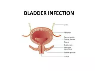
Clinical findings • Sxs hematuria: 85-90% irritative voiding Sx : CIS bone pain : bone metastasis flank pain : retroperitoneal metastasis or ureteral obstruction • Signs bimanual exam. under general anesthesia
Lab. findings • routine testing hematuria with or without pyuria azotemia, ureteral obstruction anemia • urinary cytology & flow cytometry : flow cytometry 와 cytology는 재발과 intravesical chemotherapy의 response monitoring에 이용
Imaging 목적: bladder cancer의 확진은 cystoscopy와 biopsy로 이루어짐 - evaluation of upper urinary tract - detection of muscle wall infiltration & presence of regional or distant metastasis
IVP • most common imaging test for hematuria • findings: - filling defect projecting into the lumen - fixation or flattening of bladder wall in nonpapillary, infiltrating tumor - hydronephrosis in ureteral obstruction
US • mass: echogenic foci projecting into bladder • bladder wall invasion: normal wall은 intensely echogenic 하나,less echogenic tumor로 interruption된다.
CT & MRI • bladder wall invasion과 enlarged pelvic LN detect • overall staging accuracy CT 40 - 85% MRI 50 - 90%
Cystoscopy & TUR • bladder tumor의 확진 1> superficial, low-grade tumor - single or multiple papillary - < 3cm 2> higher grade tumor - larger, sessile 3> CIS - flat area of erythema & mucosal irregularity - initial bladder tumor 의 15% 이하
Treatment Superficial bladder tumor(Ta, T1) - TUR &/or intravesical chemotherapy or immunotherapy - initial, low-grade, small tumor : TURB alone with surveillance
Agents of intravesical CTx • Thiotepa • Mitomycin-C • Adriamycin(Doxorubicin) • Epodyl • BCG: 반응율 60-80%
High Risk Group • TURB with intravesical CTx stage T1 multiple ( >4개) large ( >5cm) high grade CIS or severe dysplasia
Localized bladder tumor (T2, T3) • TURB or laser vaporization • Radical Cystectomy with/without pelvic lymphadnectomy -♂: bladder with fat, peritoneal attachment, prostate, SV -♀: bladder with fat, peritoneal attachment, cervix, uterus, ant. vaginal wall, ovary • partial cystectomy
Metastatic disease • Systemic CTx.: cisplatin based M-VAC, CMV, CISCA • External beam irradiation deeply infiltrating cancer에서 cystectomy 대신에 5,000-7,000 rad를 5-8 주에 실시 실시 bowel, bladder & rectal Cx: 15% • Chemoradiotherapy
병기별 치료법과 예후 병 기 치 료 법 5년 생존율 표재성 (65%) Ta 경요도절제 90% T1 BCG (고위험군) 71% 침윤성 (21%) T2 방광적출술 53% T3 림프절절제술 39% T4 20% 전이성 (14%) N(+), M(+) MVAC 10%이하
추적관찰 • 첫 2년: 3개월에 한번씩 Urine cytology,cystoscopy • 다음 2년은 6개월 • 다음 매년