Download
1 / 46
470 likes | 870 Views
Assessment of rectal cancer response to neoadjuvant chemoradiation – impact to clinical decision. 18 th International Gastrointestinal Cancer Conference, Istanbul, 7 th – 9 th December 2018. Ivana Bla ž i ć , MD, PhD Clinical Hospital Center Zemun Belgrade, Serbia. Rectal cancer MRI.

E N D
Assessment of rectal cancer response to neoadjuvantchemoradiation – • impact to clinical decision 18th International Gastrointestinal Cancer Conference, Istanbul, 7th – 9th December 2018 IvanaBlažić, MD, PhD Clinical Hospital Center Zemun Belgrade, Serbia
Rectal cancer MRI EURECCA. Eur. J of Cancer 2014
Rectal cancer MRI • MRI is modality of the first choice in rectal cancer diagnostic – both for primary staging and for response evaluation • standard therapy: TME with or without neoadjuvant CRT • locally advanced rectal cancers – preoperative treatment (CRT) • organ preserving treatment strategies for complete responders – ypT0
Baseline rectal cancer MRI • tumor position • tumor length • tumor morphology • T stage • circumferential resection margin (CRM) • N stage • extramural vascular invasion (EMVI)
Baseline rectal cancer MRI • tumor position • tumor length • tumor morphology • T stage • circumferential resection margin (CRM) • N stage • extramural vascular invasion (EMVI)
Baseline rectal cancer MRI • tumor position • tumor length • tumor morphology • T stage • circumferential resection margin (CRM) • N stage • extramural vascular invasion (EMVI)
Baseline rectal cancer MRI • tumor position • tumor length • tumor morphology • T stage • circumferential resection margin (CRM) • N stage • extramural vascular invasion (EMVI)
Baseline rectal cancer MRI • tumor position • tumor length • tumor morphology • T stage • circumferential resection margin (CRM) • N stage • extramural vascular invasion (EMVI) T2 T3 T4
Baseline rectal cancer MRI • tumor position • tumor length • tumor morphology • T stage • circumferential resection margin (CRM) • N stage • extramural vascular invasion (EMVI) T3 CRM – T3 CRM +
Baseline rectal cancer MRI • tumor position • tumor length • tumor morphology • T stage • circumferential resection margin (CRM) • N stage • extramural vascular invasion (EMVI) N2
Baseline rectal cancer MRI • tumor position • tumor length • tumor morphology • T stage • circumferential resection margin (CRM) • N stage • extramural vascular invasion (EMVI)
Rectal cancer MRI rectal cancer STAGING • NEOADJUVANT THERAPY DECISION • key questions: • T2 vs. T3a/b • nodal stage (N0 vs. N+) DIAGNOSIS neoadjuvant CRT THERAPY rectal cancer RESTAGING Blazic. British J Radiology 2016
Rectal cancer staging RECTAL WALL: mucosa, submucosa and lamina muscularispropria • T1 – mucosa and submucosa • T2 – lamina muscularispropria • T3 – mesorectal fat tissue • T4 – peritoneum or adjacent organs
Rectal cancer staging T1 and T2 tumors
Rectal cancer staging T3 tumors • tumor extension beyond muscularispropria: • T3a <1 mm • T3b 1-5 mm • T3c 5-15 mm • T3d > 15 mm CRM
MRI anatomy of rectum MESORECTAL FASCIA TME resection plane
Circumferential resection margin MRF involvement T3 CRM + T3 CRM –
Rectal cancer staging T2w DWI ADC T4 tumor • T4a- tumor penetrates to the surface of the visceral peritoneum • T4b - tumor directly invades other organs or structures
Rectal cancer staging CHALENGE – discrimination between T2 vs. T3a/b
Rectal cancer staging DISCRIMINATION BETWEEN T2 vs. T3 • sensitivity 82-94% • specificity 70-75% • OVERSTAGING in 29-40% • - desmoplastic reaction • UNDERSTAGING in 11% • - microscopic infiltration of mesorectal fat Bipat. Radiology 2004 Suppiah. ColorectDis 2009 Kauer. SurgEndosc 2004 Al-Sukhni. ASO 2012
Rectal cancer staging T1, T2 and T3a/b tumors (≤5mm of extramural growth) Risk of local recurrence 3% Taylor. Ann of Surg 2011
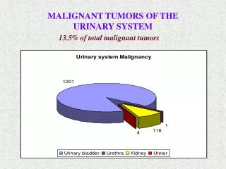
Nodal staging • measurements of LN short axis is a standard approach in LN characterization • LN size is insufficient criterion for discrimination benign from malignant LN • *high incidence of micrometastases in small LN (< 5 mm) • accurate discrimination between benign and metastatic LN is limited CHALENGE – > 50% of malignant LN is < 5 mm Perez. Dis Colon Rectum 2009 Lambregts. EurRadiol 2011 Maier. Eur J Radiology 2003
Nodal staging • detection of malignant LN based on imaging remains the crucial challenge (regardless to the modality applied) • around 25% of nodes are overstaged – unnecessary preoperative treatment and associated morbidity: • SHORT-TERM: proctitis • LONG-TERM: faecal incontinence, bowel and urogenital dysfunction • aim – to avoid over-inclusion of benign nodes Bipat. Radiology 2004
Nodal staging MRI MORPHOLOGIC PREDICTORS • heterogeneous signal intensity • irregular border • round shape Brown. Radiology 2003
Nodal staging Van Loenhout. Radiology Assistant 2015
Nodal staging EXTRAMESORECTAL LYMPH NODES • internal iliac • external iliac • obturator do not forget to evaluate extramesorectal nodes
MRI for evaluation of treatment response in rectal cancer TYPE OF TREATMENT DECISION (TME vs. organ saving treatment – NOM/TAE) key question: response evaluation rectal cancer STAGING DIAGNOSIS neoadjuvant CRT THERAPY rectal cancer RESTAGING Blazic. Radiology 2017 Sloothaak. Br J Surg 2013 West. Eur J SurgOncol 2016
MRI for evaluation of treatment response in rectal cancer NEOADJUVANT TREATMENT • long course RT (50.4 [28 x 1.8] Gy) with concomitant chemotherapy (5FU with or without additional agents) • short course RT (5x5 Gy) • induction chemotherapy followed by CRT
MRI for evaluation of treatment response in rectal cancer Restaging after CRT • tumor position – correlation with baseline MRI • yT stage • yN stage • yMRF • yEMVI
MRI for evaluation of treatment response in rectal cancer GOOD RESPONSE
MRI for evaluation of treatment response in rectal cancer beforeCRT GOOD RESPONSE after CRT
MRI for evaluation of treatment response in rectal cancer beforeCRT POOR RESPONSE after CRT
MRI for evaluation of treatment response in rectal cancer Restaging after CRT • yT stage and yMRF • normal rectal wall with no visible mass – complete response • reappearing of fat-pad – uninvolved MRF • intermediate signal tissue – residual tumor/ involved MRF • fibrotic scar tissue in tumor bed or persistent stranding around MRF CHALENGE – interpretation of post-irradiation fibrosis Blazic. British J Radiology 2016
MRI for evaluation of treatment response in rectal cancer Restaging after CRT • yN stage • nodal size is more reliable than at primary staging (NPV 81-100%) • for nodes ≤4.5-5.0 mm chance of malignancy is 3-14% • round shape + irregular border + heterogeneous signal intensity CHALENGE – discrimination between benign and metastatic LN Lahaye. Radiology 2009 Sassen. EurRadiol 2013
MRI for evaluation of treatment response in rectal cancer CLINICAL COMPLETE RESPONSE • ultimate goal of any type of neoadjuvant treatment • pCR in 8-34% of treated patients • candidates for non-operative management or organ-preserving procedures (watchful wait and transanal excision) Mass. Ann SurgOncol 2015
MRI for evaluation of treatment response in rectal cancer beforeCRT after CRT
MRI for evaluation of treatment response in rectal cancer DWI ? T2w ADC
MRI for evaluation of treatment response in rectal cancer DWI • DWI is recommended for restaging, mainly for yT staging • enables qualitative and quantitative assessment of tumor response • improves MRI accuracy for assessment of CR Paardtet. Radiology 2013
MRI for evaluation of treatment response in rectal cancer ADVANCED TECHNIQUES • combination of clinical examination (DRE and endoscopy) with MRI/DWI(probability for prediction CR of 98%) for accurate response evaluation • insufficient evidence to support use of DCE MRI for restaging • radiomics analysis can be used for rectal cancer response evaluation Blazic. Radiology 2017 Maas. Ann SurgOncol 2015 Gollub, Blazic. EurRadiol 2018 Horvat, Blazic. Radiology 2018
Rectal cancer MRI Rectal cancer staging • assessment of T stage, N stage, MRF and EMVI • neoadjuvant treatment decision • CHALENGES • discrimination between T2 vs. T3a/b • discrimination between benign and metastatic lymph nodes FUTURE PERSPECTIVES avoid overcalling of positive nodes
Rectal cancer MRI Restaging after CRT • reassessment of yT stage, yN stage, yMRF and yEMVI • type of treatment decision (TME vs. organ saving treatment – NOM/TAE) • CHALENGES • interpretation of post-irradiation fibrosis • discrimination between benign and metastatic lymph nodes • nodal restaging is more accurate than primary staging
Rectal cancer MRI Restaging after CRT • post-treatment MRI has important impact on treatment choice • endoscopy and digital exam remain main assessment tools • MRI with DWI plays key role among imaging methods • all three combined reach the highest accuracy OPTIMAL STRATEGY combination of clinical examination with MRI/DWI
Assessment of rectal cancer response to neoadjuvantchemoradiation – • impact to clinical decision 18th International Gastrointestinal Cancer Conference, Istanbul, 7th – 9th December 2018 IvanaBlažić, MD, PhD Clinical Hospital Center Zemun Belgrade, Serbia