
Angina pectoris 狭心症
E N D
Presentation Transcript
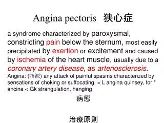
Angina pectoris 狭心症 a syndrome characterized by paroxysmal, constricting pain below the sternum, most easily precipitated by exertionor excitement and caused by ischemia of the heart muscle, usually due to acoronary artery disease, as arteriosclerosis.Angina: (語源) any attack of painful spasms characterized by sensations of choking or suffocating. < L angina quinsey, for * ancina < Gk strangulation, hanging 病態 治療原則
男性、中年 季節:冬 寒冷暴露 次の有名な絵を見て, 狭心症の発作につながる 因子を挙げよ 大食後 重い荷物 階段登り タバコ 前胸部痛
収縮力 冠動静脈酸素含有量の較差 酸素 血圧(後負荷) 酸素 大動脈圧 心拍数 供給 需要 (拡張期) 心筋酸素分布 冠血流量 左心室圧 冠血管抵抗 壁張力 心室容積 静脈還流(前負荷) 虚血 自覚症状 心電図異常 代謝障害 心室機能不全 乳酸・H+ の産生 壁運動異常 ST下降(労作性狭心症) + 拡張終期圧上昇 ST上昇(安静時狭心症) K の漏出 不整脈 心拍出量低下 アデノシン遊離 図4-2 心筋酸素需要・供給と狭心症の病態 狭心痛
狭心痛の 部位と放散
心拍出量の分配率と酸素消費 動脈血には20 vol% の酸素が含まれている O2 extraction vol% CaO2 – CvO2 心臓11.5%,脳6.4、肝臓4.1、腎臓1.3、 骨格筋静止時4.3、最大運動時17.2
Lead V4 at rest (top) and after 4.5 min of exercise (bottom). There is 0.3 mV of horizontal ST-segment depression, indicating a positive test for ischemia.
Coronary Angiography Stenosis: narrowing of the artery, due most likely to atherosclerosis. Damage to the intima due to atherosclerosis is a major cause of thrombosis in the arteries. The resultant ischemia of the myocardium can lead to ischemic necrosis, i.e., an infarct [Myocardial Infarction]. • What therapeutic agent can be used to lyse the clots in coronary vessels? • How do the various natural anticoagulants act?
冠動脈硬化 Coronary atherosclerosis 内膜下のプラーク生成 血栓生成の可能性 Spasm発生の可能性
冠動脈血栓 Coronary thrombosis 内皮細胞機能 血小板 マクロファージ 増殖型平滑筋細胞 図2-20参照、さらに病理学で学んでください。
Dynamic coronary obstruction tone tone Spasm Rest angina
Dynamic coronary obstruction • This figure shows how the caliber of eccentric coronary artery stenoses may change, with considerable variation in the degree of stenosis resistance and the propensity to produce angina. • Both increased vascular tone (first two examples, spasm & tone ) and decreased vascular tone (third example) are depicted.
狭心症 angina pectoris の分類 (p.139) 安定狭心症 Stable (typical) angina 器質性(労作性) プリンツメタル型狭心症 Prinzmetal (variant) angina 冠動脈攣縮型(異型) 不安定狭心症 Unstable (crescendo) angina 梗塞前
安定狭心症 ・安定プラークによる冠状動脈 (LAD,LCX,RCA)の内腔狭窄 『酸素需要>酸素供給』→休息により改善
冠動脈攣縮 spasm 血管平滑筋の過剰収縮 冠血管狭窄 冠血流量減少
プリンツメタル型狭心症 • 冠状動脈のspasm (攣縮)による • (plaque プラークの近傍) • 休息・睡眠時 • ←運動・血圧・心拍数に無関係 • 喫煙がリスクファクター • より若年で発症。予後比較的良好。 • ST-segment elevation ←貫壁性の虚血
不安定狭心症 (Preinfarction angina) ・不安定プラークの崩壊 (アテローム内出血、機械的ストレス↑) →血栓形成・Vasospasm→一時的内腔閉塞 ・心筋梗塞へ移行しやすい →動脈硬化病変が不安定であり、 続発性の血栓形成が起こりやすい ・頻度が増大、発作の延長。休息時にも起こる。
動脈硬化から 見た狭心症
閉塞性血栓 1 白色血栓症 動脈壁の異常 血小板とフィブリンが主役
閉塞性血栓 2 赤色血栓症 プラークの破綻 凝固因子が主役
図4-1冠動脈硬化およびスパスムと狭心症治療薬の位置付け図4-1冠動脈硬化およびスパスムと狭心症治療薬の位置付け 抗脂血症 治療薬
Correction of the imbalance increasing delivery (by increasing coronary flow) nitrates (nitroglycerin) vasodilators (Ca antagonists, KCOs) coronary bypass grafts or angioplasty (PTCA) percutaneous transcatheter coronary angioplasty decreasing oxygen demand (by decreasing cardiac work) b-blockes, nitrates nitrates (nitroglycerin) vasodilators (Ca antagonists, KCOs) *against atherosclerosis and thrombus formation
収縮力 冠動静脈酸素含有量の較差 酸素 血圧(後負荷) 酸素 大動脈圧 心拍数 供給 需要 (拡張期) 心筋酸素分布 冠血流量 左心室圧 冠血管抵抗 壁張力 心室容積 静脈還流(前負荷) 虚血 自覚症状 心電図異常 代謝障害 心室機能不全 + 乳酸・H の産生 壁運動異常 狭心痛 ST下降(労作性狭心症) + 拡張終期圧上昇 ST上昇(安静時狭心症) K の漏出 不整脈 心拍出量低下 アデノシン遊離 図4-2 心筋酸素需要・供給と狭心症の病態 small oxygen debt: extract about 75% of available oxygen ‘tachycardia’-- harmful
冠状動脈の分岐(A)と血流量変化(B). 心周期中に冠状動脈の血流は変化する。 2本の点線の 間は心室の拍出期で、大動脈圧は高いが、左心室の内圧がさらに高いために左冠状動脈の血流は減少している。(図15-24,生理学テキスト) 時間 (秒)
Artery blocked by thrombus on fissured atherosclerotic plaque Zone of perfusion (area at risk) next
Myocardium 24 hr 2 hr 0 hr Obstructed coronary art. Endocardium Zone of necrosis Zone of necrosis area at risk Epicardium Progression of myocardial necrosisafter coronary artery occlusion. Necrosis begins in a small zone of the myocardium beneath the endocardial surface in the center of the ischemic zone. This entire region of myocardium (dashed outline) depends on the occluded vessel for perfusion and is the area at risk. Note that a very narrow zone of myocardium immediately beneath the endocardium is spared from necrosis because it can be oxygenated by diffusion from the ventricle. (Fig. 13-6 in Robbins)
Principle of stress radionuclide imaging for the detection of coronary artery disease. • a normal coronary artery and an artery with significant stenosis (top), • (2) the myocardial territory (perfusion image), and • (3) the ventricular wall (wall motion image) supplied by each artery.
Distribution of regional myocardial blood flow • The distribution of regional myocardial blood flow in the myocardium is heterogeneous, i.e., there is less blood flowing in the ischemic myocardial bed (blue area) than in the normal myocardial bed.
Coronary blood flow at rest and during stress (x ~5) 最大運動時
Coronary blood flow at rest and during stress (1) At rest, coronary blood flow is similar in the normal artery and in the artery with stenosis. Resting coronary blood flow in the diseased artery is maintained at a normal level and is sufficient to meet resting metabolic myocardial demands because of recruitment of coronary reserve (dilatation of the resistance vasculature) in the distal coronary bed. During exercise, myocardial metabolic demands increase and more myocardial nutrient blood flow is required. Coronary reserve is increased as a result of dilatation of the coronary resistance vessels in the peripheral coronary bed.
Coronary blood flow at rest and during stress (2) In the territory of the normal coronary artery, the resistance vessels dilate and coronary blood flow is increased by 2 to 2.5 times. In the abnormal coronary bed, which is distal to the significant coronary stenosis, resistance vessels are already dilated and little if any further dilatation is possible. Increased metabolic demands thus cannot be met, and the relatively hypoperfused myocardium becomes ischemic.