Download
1 / 16
190 likes | 570 Views
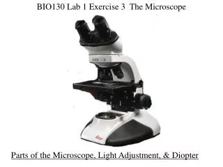
BIO130 Lab 1 Exercise 3 The Microscope. Parts of the Microscope, Light Adjustment, & Diopter. Measuring Field Diameter Using the Stage Micrometer: 1. Insure that left ocular lens is turned fully counterclockwise (Lens should be as tall as it can be) 2. Focus on the stage micrometer

E N D
BIO130 Lab 1 Exercise 3 The Microscope Parts of the Microscope, Light Adjustment, & Diopter
Measuring Field Diameter Using the Stage Micrometer: 1. Insure that left ocular lens is turned fully counterclockwise (Lens should be as tall as it can be) 2. Focus on the stage micrometer 3. Measure the diameter (widest part) of the field of view using the tiny millimeter ruler of the stage micrometer using only your left eye in the left ocular 40X Magnification
100X Magnification Read off where the ruler ends on the other side: this one looks like 1.745mm Each scope is a little different Line up the 0.0 line on the edge of the visible field: the zeros will no longer be visible
Using field diameter to determine the size of an object specimen feature or cell: 1. Know the field diameter of the objective lens you are looking through. (We measured this) 2. Center and focus the object. 3. “Guess” how many of that object would fit end to end across the diameter of the field. (Diameter = the widest part of the circle of light you see through the lens) 10X objective: diameter measured to be 1860µm Looks like 2 would fit across 1860µm
Using field diameter to determine the size of an object specimen feature or cell: 4. Divide the field diameter by the number of objects you think would fit across end to end to determine the size of one of the objects. 10X objective: diameter measured to be 1860µm Looks like 2 would fit across 1860µm ÷ 2 = 930µm 1860µm
New object: 10X objective: diameter measured to be 1860µm Looks like 4 would fit across 1860µm 1860µm ÷ 4 = 465µm The green oval is approximately 465µm long
Which lens do I use? The object is a fixed size, regardless of lens you should get the same answer! Use the lens that allows you to see the whole object the best: big enough to guestimate how many fit across the diameter, not too magnified that you cannot see the whole object in the same field.
10X objective: diameter measured to be 1860µm Looks like 4 would fit across 1860µm ÷ 4 = 465µm 1860µm 40X objective: diameter measured to be 465µm Looks like 1 would fit across 465µm ÷ 1 = 465µm 465µm This object (green oval) is 465µm: you should get approximately the same answer which ever lens you use.
4X objective (40X) Dogfish Placoid Scale Measure one diamond 10X objective (100X) 40X objective (400X)
http://www.elasmodiver.com/Sharkive%20images/Spiny-dogfish-070.jpghttp://www.elasmodiver.com/Sharkive%20images/Spiny-dogfish-070.jpg http://relentlessthirst.files.wordpress.com/2009/02/dogfish1.jpg
4X objective (40X) Dinoflagellate Don’t cut off his tails! 10X objective (100X) 40X objective (400X)
Red Tide http://www.sciencedaily.com/images/2007/08/070830150106-large.jpg http://www.pac.dfo-mpo.gc.ca/fm-gp/contamination/Images/Red%20Tide%20-%20Mary%20Mackin%202.jpg
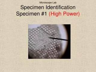
4X objective (40X) Human Cheek Cell Large irregular shaped cells: look like a ruffled sunny side up egg 10X objective (100X) 40X objective (400X)
4X objective (40X) Human Blood Smear Tiny red dots, even with the 40X objective: guestimate carefully! 10X objective (100X) 40X objective (400X) WBC RBC
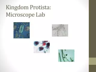
Dogfish Placoid Scale Dinoflagellate Human Cheek Cell Human Blood Smear WBC RBC