Download
1 / 19
190 likes | 430 Views
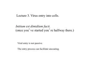
Lecture 3. Virus entry into cells. Initium est dimidium facti. (once you ’ ve started you ’ re halfway there.). Viral entry is not passive. The entry process can facilitate uncoating. Plasma membrane showing integral and peripheral membrane proteins. Many proteins on the outside of cell

E N D
Lecture 3. Virus entry into cells. Initium est dimidium facti. (once you’ve started you’re halfway there.) Viral entry is not passive. The entry process can facilitate uncoating.
Plasma membrane showing integral and peripheral membrane proteins. Many proteins on the outside of cell are glycoproteins Figure 4.4 Virology Note how impermeable the membrane is. Viruses cannot pass freely through membrane (in either direction). Many virus receptors are proteins (often glycoproteins) on the host cell
Mechanisms for uptake of macromolecules from extracellular fluid. Uptake by macrophages Figure 5.1 Virology Size of viruses Large particles are engulfed by a phagocytic cell, such as a macrophage. Extensions fuse around the particle. Phagosomes fuse with lysosomes and the particles are destroyed. Macrophages use phagocytosis to take up bacteria and viruses. Most important mechanism of entry for viruses is receptor-mediated endocytosis.
Receptor mediated endocytosis 1. Ligand binds a cell surface receptor. This diffuses into an invagination coated with clathrin. 2. The clathrin pit pinches off forming a 'coated vesicle'. 3. The clathrin uncoats the vesicle. Figure 5.2 Virology 4. The vesicle fuses with the early endosome. The early endosome is acidic. 5. The acidification releases the ligand from the receptor, and receptor is returned to cell surface.
Viruses have evolved to use proteins on the host cell as receptors. Rhinovirus and poliovirus are related picornaviruses. Icam-1 Rhinovirus receptor Pvr-poliovirus receptor Rhinoviruses cause the common cold. More than 90 related types. Icam-1 is involved in Macrophage T-cell interactions. PVR is involved in DC-NK interactions Figure 4.9 Virology Intercellular adhesion molecule-1 Presence of the receptor determines specificity of virus host range (tropism). for example mice do not have Pvr and are not infected with poliovirus. Human and Mouse ICAM-1 are different enough that human rhinoviruses cannot infect mice.
The domain 1 of Icam-1 binds deep within the ‘canyon’ of rhinovirus Direct interaction between receptor and capsid. Why is it significant that the receptor binding site is recessed? Figure 4.14 Virology
Depth view of the surface of poliovirus White-farthest from center Black-nearest center canyon Hogle J. 2002 Annu. Rev. Microbiol. 56:677
Binding sites for LFA-1 and human rhinovirus (HRV) overlap Icam-1 HRV LFA-1 Why don’t we mutate our ICAM-1 to not allow binding to rhinovirus? Figure 4.10 Virology
Human rhinovirus entry and uncoating. The virus attaches to Icam-1 and enters by endocytosis. The acidic environment of the endosome causes the uncoating of the particle. The RNA is released into the cytoplasm. Icam-1 deep in canyon facilitates destabilization of capsid. Rhinovirus is not resistant to acid, unlike poliovirus. Figure 14.19 Virology
Neutralizing antibody blocks binding of the receptor to the canyon Figure 4.14 Virology
Models of poliovirus entering the cell Pvr interaction causes major structural changes. Poliovirus is acid- resistant. Figure 5.13 Virology
Formation of a pore in the membrane by poliovirus Figure 5.13 Virology Interaction with Pvr causes a major structural change. The N termini of the VP1 protein extends into the membrane. This may form a pore in which the RNA can enter. Fc-receptor mediated internalization of poliovirus (say by a macrophage) does not allow uncoating.
An enveloped virus also has to escape from the membrane Figure 5.3 Virology Membrane fusion is a highly regulated process involving proteins for targeting and docking. Enveloped viruses encode their own ‘fusogens’.
Sialic acid is the cellular receptor for influenza virus attachment Integral membrane glycoproteins are the receptors for influenza binding. Figure 4.11 Virology
Influenza virus hemagglutinin (HA) binds to terminal sialic acid. Galactose chain An a (2,3) glycosidic linkage is shown (preferred by avian HA). Human influenza HA prefers to bind to an a (2,6) linked sialic acid.
Influenza virus entry illustrating the HA fusion Fusogenic peptide is exposed by an acid catalyzed structural change Virus nucleic acid is inside two membranes. To it get out it uses its fusogen Acidic environment of the endosome causes a conformational change in HA Figure 5.7 Virology
Conformational changes in HA2 to expose fusogenic peptide Ann. Rev. Biochem. 2000. 69: 531
Influenza hemagglutinin conformational change exposing the fusogenic peptide. Entire HA molecule HA2 half only shown Ann. Rev. Biochem. 2000. 69: 531
Further structural changes in HA induce membrane fusion Ann. Rev. Biochem. 2000. 69: 531