Clinical Metrics of Resolved Cornea vs. Cornea with MGK Development: Findings and Observations
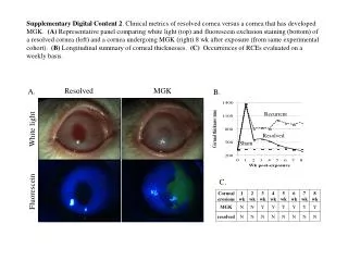
This document presents clinical metrics comparing a resolved cornea to one that has developed meibomian gland keratinization (MGK). Part A features representative panels showcasing differences observed in white light and fluorescein exclusion staining between the two corneas eight weeks post-exposure. Part B offers a longitudinal summary of corneal thickness measurements, while Part C evaluates the occurrences of recurrent corneal erosions (RCEs) on a weekly basis within the same experimental cohort. These findings contribute valuable insights into corneal health and disease.

Clinical Metrics of Resolved Cornea vs. Cornea with MGK Development: Findings and Observations
E N D
Presentation Transcript
Supplementary Digital Content 2. Clinical metrics of resolved cornea versus a cornea that has developed MGK. (A) Representative panel comparing white light (top) and fluorescein exclusion staining (bottom) of a resolved cornea (left) and a cornea undergoing MGK (right) 8 wk after exposure (from same experimental cohort). (B) Longitudinal summary of corneal thicknesses. (C) Occurrences of RCEs evaluated on a weekly basis. Resolved MGK A. B. Recurrent White light Resolved Sham C. Fluorescein