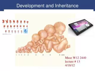
Development and Inheritance
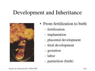
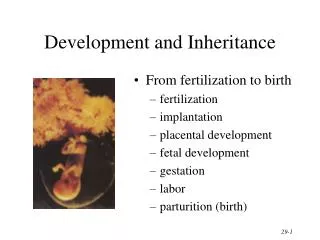
Development and Inheritance. From fertilization to birth fertilization implantation placental development fetal development gestation labor parturition (birth). Terminology of Development. Gestation period time span from fertilization to birth (38 weeks) Prenatal period (before birth)

Development and Inheritance
E N D
Presentation Transcript
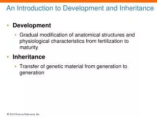
Development and Inheritance • From fertilization to birth • fertilization • implantation • placental development • fetal development • gestation • labor • parturition (birth)
Terminology of Development • Gestation period • time span from fertilization to birth (38 weeks) • Prenatal period (before birth) • embryological development • developing human for first 2 months after fertilization is known as an embryo • all principal adult organs are present • fetal development • from 9 weeks until birth is known as a fetus • by end of 3rd month, placenta is functioning • Neonatal period is first 42 days after birth • obstetrics is branch of medicine that deals with the neonatal period, pregnancy and labor
Events Before Fertilization • Haploid sperm nucleus & haploid secondary oocyte nucleus merge to form a single diploid nucleus • Occurs in uterine tube within 24 hours after ovulation (oocyte dies in 24 hours) • Events occurring before fertilization • peristalsis of uterine tube & movement of cilia transport the oocyte towards the uterus • oocyte releases chemical attractants • sperm swim towards oocyte by means of flagella • prostaglandins within the semen stimulate uterine contractions that help move sperm towards the oocyte • capacitationor final maturation of the sperm occurs within female • acrosomal membrane becomes fragile
Sperm Contact during Fertilization • Sperm penetrates the granulosa cellsaround the oocyte (corona radiata) • Sperm digests its way throughthe zona pellucida • when ZP3 glycoprotein binds to spermhead, it triggers the acrosomal reaction(enzyme release) • First sperm to fuse with oocyte membrane triggers the slow & the fast block to polyspermy • 1-3 seconds after contact, oocyte membrane depolarizes & other cells can not fuse with it = fast block to polyspermy • depolarization triggers the intracellular release of Ca+2 causing the exocytosis of molecules hardening the entire zona pellucida = slow block to polyspermy
Events Within the Egg • Sperm entry, triggers oocyte to complete meiosis II and dump second polar body • Once inside the oocyte, the sperm loses its tail & becomes a male pronucleus • Fusion of male & female haploid pronuclei is the true moment of fertilization • Fertilized ovum (2n) is called a zygote • zona pellucida still surrounds it
Nature of Twins • Fraternal twins (dizygotic) • independent release of 2 oocytes fertilized by 2 separate sperm • as different as any 2 siblings • Identical twins (monozygotic) • 2 individuals that develop from a single fertilized ovum • genetically identical & always the same sex • if ovum does not completely separate, conjoined twins (share some body structures)
Formation of the Morula • Rapid mitotic cell division of embryo is called cleavage • 1st cleavage in 30 hours produces 2 blastomeres • 2nd cleavage on 2nd day • By 3rd day has 16 cells • By day 4 has formed a solidball of cells called a morula
Development of the Blastocyst • A blastocyst is a hollow ball of cells that enters the uterine cavity by day 5 • outer covering of cellscalled the trophoblast • inner cell mass • fluid-filled cavity calledthe blastocele • Trophoblast & part of innercell mass will develop into fetal portion of placenta • Most of inner cell mass will become embryo
Implantation • Attachment of blastocyst to endometrium • occurs 6 days after fertilization • implants with inner cell mass in contact with the endometrium • Trophoblast develops 2 distinct layers • syncytiotrophoblast secretes enzymes that digest the endometrial cells • cytotrophoblast is distinct layer of cells that defines the original shape of the embryo • Trophoblast secretes human chorionic gonadotropin (hCG) that helps the corpus luteum maintain the uterine lining
Implantation Notice: distinct syncytiotrophoblast and cytotrophoblast layers.
Ectopic Pregnancy • Development of an embryo outside the uterus • Most often in uterine tube • common causes are blockages of uterine tube such as tumors or scars from pelvic inflammatory disease • symptoms are missed menstrual cycles, bleeding & acute pain • Twice as common in smokers because nicotine paralyzes the cilia
Beginnings of Organ Systems(Gastrulation) • Day 8 • cytotrophoblast forms amnion & amnionic cavity • cells of inner cell mass on amnionic cavity form ectoderm • cells bordering on blastocele form endoderm • ectoderm & endoderm together form embryonic disk • Day 12 • endodermal cells divideto form a hollow sphere(yolk sac) • cytotrophoblast cellsdivide to fill the spacessurrounding the yolk sac with extraembryonic mesoderm • spaces develop in that layer to form future ventral body cavity
Primary Germ Layers • Day 14 --cells of embryonic disc produce 3 distinct layers • endoderm forms epithelial lining of GI & respiratory • mesoderm forms muscle, bone & other connective tissues • ectoderm develops into epidermis of skin & nervous system
Formation of Embryonic Membranes • Yolk sac • site of early blood formation • gives rise to gonadal stem cells (spermatogonia & oogonia) • Amnion • surrounds embryo with fluid: shock absorber, regulates body temperature & prevents adhesions • fluid is filtrate of mother’s blood + fetal urine • examine a sample of it for embryonic cells (amniocentesis) • Chorion • becomes the embryonic contribution to the placenta • derived from trophoblast & mesoderm lining it • gives rise to human chorionic gonadotropin (hCG) • Allantois • outpocketing off yolk sac that becomes umbilical cord
Placenta & Umbilical Cord • Placenta forms during 3rd month • chorion of embryo & stratum functionalis layer of uterus • Chorionic villi extend into maternal blood filled intervillous spaces --- maternal & fetal blood vessels do not join & blood does not mix • diffusion of O2, nutrients, wastes • stores nutrients & produces hormones • barrier to microorganisms, except some viruses • AIDS, measles, chickenpox, poliomyelitis, encephalitis • not a barrier to drugs such as alcohol • Placenta detaches from the uterus (afterbirth)
Parts of Endometrial Lining • Decidua = all of endometrium lost as placenta • equals all of the endometrium, except stratum basalis • Decidua basalis---portion ofendometrium deep to chorion • Decidua capsularis---part ofendometrial wall that coversimplanted embryo • Decidua parietalis---part ofendometrial wall not modifiedby embryo until embryo bumps into it as it enlarges • Decidua capsularis fuses with decidua parietalis
Umbilical Cord • Contents • 2 arteries that carry blood to the placenta • 1 umbilical vein that carries oxygenated blood to the fetus • primitive connective tissue • Stub drops off in 2 weeks leaving a scar called the umbilicus
Placenta Previa • Placenta is implanted near or covering os of cervix • occurs in 1 to 250 live births • May lead to spontaneous abortion, premature birth or increased maternal mortality • Major symptom is sudden, painless bright red vaginal bleeding in the 3rd trimester • Cesarean section is preferred delivery method
Fetal Ultrasonography • Transducer emits high-frequency sound waves • reflected sound waves converted to on-screen image called sonogram • patient needs full bladder • Used to determine fetal age, viability, growth, position, twins and maternal abnormalities
Amniocentesis • Fetal cells from 10 ml sample of amniotic fluid examined for genetic defects • Test at 14-16 weeks • Results back in one month • Needle through abdominal wall & uterus • Chance of spontaneous abortion is 0.5%
Chorionic Villi Sampling • As early as 8 weeks • Results in few days • Chance of spontaneous abortion 1-2% • 30 mg of placenta removed by suctionthrough cervix or with needle through abdomen • Chromosomal analysis reveals same results as amniocentesis
Hormones of Pregnancy • Chorion • from day 8 until 4 months secretes hCG which keeps corpus luteum active • corpus luteum produces progesterone & estrogen to maintain lining of uterus • Placenta • by 4th month produces enough progesterone & estrogen that corpus luteum is no longer important • relaxin which relaxes CT of pelvis and cervix • human chorionic somatomammotropoin (hCS) or human placental lactogen (hPL) • maximum amount by 32 weeks • helps prepare mammary glands for lactation • corticotropin-releasing hormone (CRH) increases secretion of fetal cortisol (lung maturation) & acts to establish timing of birth
Hormone Blood Levels • Human chorionic gonadotropin (hCG) produced by the chorion is less important after 4 months, because the placenta takes over the hormonal secretion of the corpus luteum.
Early Pregnancy Tests • Detect human chorionic gonadotropin (hCG) in the urine as soon as 8 days after fertilization • color change hen reaction between urine & antibodies in kit • False-negatives & false-positives do occur • excess protein or blood in urine • rare type of uterine cancer • steroid, diuretics, hormones and thyroid drugs alter test results
Developmental Changes • Read Table 29.2 to get a full description of the timing of fetal events during development
Maternal Changes During Pregnancy • Uterus nearly fills the abdominal cavity • GI tract compressed causing heartburn & constipation • Pressure on bladder causing changes in frequency & urgency • Compression of vena cava causing varicose veins & edema in the legs • Compression of renal vessels causing renal hypertension
Changes During Pregnancy • Cardiovascular changes to meet needs of fetus • rise in cardiac output of 20-30% due to placenta • increase in heart rate 15% & increase in blood volume 30-50% • Respiratory changes • increase in tidal volume 30% • decrease in expiratory reserve volume & airway resistance • minute respiratory volume increases as O2 needs increase • Reproductive system changes • uterus increases in size from 80 g to 1200g • hyperplasia and hypertrophy • Urinary system changes • increase in glomerular filtration rate of 40%
Pregnancy-Induced Hypertension • Elevated blood pressure • Major cause is preeclampsia • sudden hypertension • large amounts of protein in the urine • generalized edema, blurred vision & headaches • Autoimmune or allergic reaction to presence of fetus • Eclampsia = convulsions & coma in mother
Exercise and Pregnancy • In early pregnancy • avoid excessive exercise & heat buildup • linked to neural tube defects • Moderate exercise has beneficial effects • no evidence of inadequate blood flow to the placenta
Labor and Parturition • Parturition means giving birth; labor is the process of expelling the fetus • Labor begins when progesterone’s inhibition is overcome by an increase in the levels of estrogen • progesterone inhibits uterine contraction • placenta stimulates fetal anterior pituitary which causes fetal adrenal gland to secrete DHEA • placenta converts DHEA to estrogen • estrogen overcomes progesterone and labor begins
Positive Feedback during Labor • Uterine contraction forces fetal head into cervix (stretch) • Nerve impulses reach hypothalamus causing release of oxytocin • Oxytocin causes more contractions producing more stretch of cervix & more nerve impulses
True Versus False Labor • True labor begins when contractions occur at regular intervals • produces pain • back pain increases with walking • dilation of cervix with a discharge of blood-containing mucus in the cervical canal • False labor produces pain at irregular intervals but there is no cervical dilation
Stages of Labor • Dilation • 6 to 12 hours • rupture of amniotic sac & dilation of cervix • Expulsion • 10 minutes to several hours • baby moves through birth canal • Placental • 30 minutes • afterbirth is expelled by muscular contractions
Dystocia & Cesarean Section • Dystocia = difficult labor • due to fetal position or size • breech presentation is butt or feet first in birth canal • Cesarean section (C-section) • horizontal incision through lower abdominal wall and uterus • a history of multiple cesarean sections does not preclude a vaginal birth
Adjustments of the Infant at Birth • Respiratory System • after cord is cut, increased CO2 levels in blood cause muscular contractions and first breath • breathing rate begins at 45/minute for the first 2 weeks & declines to reach normal rate • Cardiovascular System • foramen ovale closes at moment of birth • ductus arteriosus & umbilical vein close down by muscle contractions & become ligaments • pulse rate slows down (120 to 160 at birth) • increase in rate of RBC & hemoglobin formation
Premature Infants • Preemie is any baby weighs less than 5lb. 8oz at birth • Causes • poor prenatal care • drug abuse • young or old mother (below 16 or above 35) • Below 36 weeks • respiratory distress syndrome due to insufficient surfactant is major problem
Physiology of Lactation • Lactation = production & release of milk • Prolactin from anterior pituitary increases during pregnancy, but progesterone inhibits effects of prolactin until after delivery • After delivery, progesterone levels drop & suckling increases the release of prolactin & oxytocin (milk ejection reflex) • Colostrum = cloudy fluid released for few days • True milk produced by 4th day • If suckling stops, milk secretion stops
Milk Ejection Reflex • Oxytocin cause release of milk into mammary ducts • Stimulation of touching nipple causes hypothalamus to release oxytocin • Oxytocin causes contraction of myoepithelial cells • Milk moved from alveoli into mammary ducts • Oxytocin release by other stimuli • hearing a baby’s cry or touching the genitals
Benefits of Breast-feeding • Faster & better absorption of the “right” nutrients • Beneficial cells • functional white blood cells • neutrophils help ingest bacteria in baby’s gut • macrophages produce lysozymes • plasma cells provides antibodies prevent gastroenteritis • Decreased incidence of diseases later in life • reduction in allergies, respiratory & GI infections, ear infections & diarrhea • Parent-child bonding • Infant in control of intake
Nursing and Childbirth • Nursing of first-born twin speeds birth of second child • stimulates release of oxytocin • Nursing of only child • promotes expulsion of the placenta • helps control hemorrhage after birth • helps uterus return to normal size
Inheritance • Passing of hereditary traits from one generation to the next • Genotype • all human cells contain 23 pairs of chromosomes • one chromosome in each pair came from the mother and the other came from the father • similar locations on each pair of chromosomes code for the same trait (alleles) • if one allele controls the express of a trait, it is the dominant allele • if the other allele is completely masked it is the recessive allele • a person with the same alleles on both chromosomes is said to be homozygous for the trait----heterozygous for the trait is having different alleles on homologous chromosomes • heterozygous individuals are carriers of a recessive gene
Genotype & Phenotype • Genotype = your genetic makeup • Phenotype = what you look like (outward expression of your genes) • Punnett square • method of showing 4 possible genetic combinations in offspringof 2 individuals
Genetic Problems • Error in meiosis called nondisjunction • chromosomes fail to separate properly • cell with one or more extra or missing chromosomes is called an aneuploid • (2n-1) is missing a chromosome • (2n+1) has an extra chromosome • Error in meiosis called translocation • location of chromosome segment is moved • crossing-over between 2 nonhomologous chromosomes • Down syndrome results from a portion of chromosome 21 becoming part of another chromosome • individuals have 3 copies of that part of chromosome 21
Incomplete Dominance • Neither member of an allelic pair is dominant over the other --- resulting phenotype is intermediate • Sickle-cell trait individuals have both HbA & HbS • suffer from only minor problems with anemia since have both normal & sickle-cell hemoglobin • Sickle-cell anemic individuals have 2HbS alleles • produce sickle-cell hemoglobin • suffer from severe anemia
Sickle-Cell Inheritance • 1 normal • 2 embryos will be sickle-cell trait • 1 sickle-cell anemia
Multiple-Allele Inheritance • Genes with more than two alternate forms • 3 different alleles of the I gene • IA, IB, or i • A and B alleles are codominant since both genes are expressed equally • 6 possible genotypes produce 4 blood types • 4 phenotypes of the ABO blood groups are (A, B, AB & O)