Oxidation-Reduction Reactions
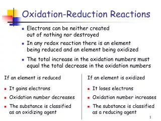
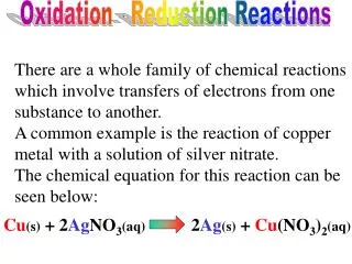
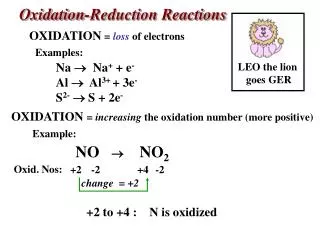
Oxidation-Reduction Reactions. In every oxidation-reduction reaction there must be an electron donor and an electron acceptor, with electrons transferred from donor to acceptor In biological systems the energy released during these oxidations is coupled to the formation of energy-rich molecules

Oxidation-Reduction Reactions
E N D
Presentation Transcript
Oxidation-Reduction Reactions In every oxidation-reduction reaction there must be an electron donor and an electron acceptor, with electrons transferred from donor to acceptor In biological systems the energy released during these oxidations is coupled to the formation of energy-rich molecules How do enzymes catalyze these electron transfer reactions ?
Oxidation States at Carbon Most enzyme-catalyzed redox processes involve the transfer of two electrons This can be accomplished either by a single two-electron step or by consecutive one-electron steps
Electron Transfers one electron steps Electron transfers can occur as a series of one-electron steps if any of the following conditions are present: A resonance-stabilized radical intermediate makes the one-electron transfer energetically favorable
Electron Transfers one electron steps Unless one of these three conditions is present we can safely assume that the electron transfer takes place in a single two-electron step
Electron Transfers two electron step Each of these electron transfer mechanisms leads to the same products Unless some additional information is obtained it is not possible to determine which of these is the correct mechanism
Pyridine-linked Dehydrogenases two electron hydride transfer reactions
Pyridine-linked Dehydrogenases coenzyme The nicotinamide ring is the hydride ion acceptor * Hydride transfer to carbon-4 produces the reduced form of the coenzyme The addition of a 2’-phosphate changes NAD to NADP
Alcohol Dehydrogenase coenzyme binding There are numerous interactions between each region of NAD(P) and active site functional groups These interactions dictate the absolute specificity for pyridine nucleotide binding The nature of the interactions near the 2’-position determine NAD vs. NADP specificity In this case the 2’-hydroxyl group of NAD is involved as both a hydrogen bond donor and acceptor
Alcohol Dehydrogenase structure the metal ion is bound adjacent to the alcohol binding site Cd(II)-form of the liver enzyme J. Biol. Chem. 276, 9316 (2000)
Alcohol Dehydrogenase active site hydrogen bonding network Two Cys and a His form the metal binding site Zinc coordinates the alcohol Ser48 is the active site base that removes the alcohol proton His51 assists by deprotonating Ser48 Hydride is transferred to the C4 of NAD Biochemistry33, 5230 (1994)
Alcohol Dehydrogenase active site of drosophila enzyme An unusual alcohol dehydrogenase that is missing the active site Zn Lys155 lowers the pK of the hydroxyl group of Tyr151 as NAD binds Ser138 aids in alcohol binding Tyr151 is the catalytic base that abstracts the proton Hydride transfer leads to the oxidized ketone and NADH products J. Molec. Biol.282, 383 (1998)
Isocitrate Dehydrogenase active site structure Ca(II)-form of the enzyme the metal ion is essential, but is only involved in one part of the catalytic cycle The metal ion site is adjacent to the substrate binding site J. Biol. Chem. 279, 33946 (2004)
Isocitrate Dehydrogenase active site structure Isocitrate is bound by interactions with three arginines (R101, R110 & R133) The carboxyl group that is removed is hydrogen-bonded to Y140 & K212 Three aspartates (D252, D275 & D279) form the divalent metal ion site What is the role of S95 (S113) ? This view shows the charged surface at the active site The next view focuses on the substrate binding groups
Isocitrate Dehydrogenase Role of Ser113 Maximum velocity (kcat) and Km for isocitrate with ICDH mutants kcat (%) 2.7% 4.8% 0.04% 1.2% 0.07% 0.4% 4.9% 100% 3.1% 1.0% Each substitution at position 113 results in a loss of catalytic activity and an increase in the Km for isocitrate What role does Ser113 play in this reaction ?
Isocitrate Dehydrogenase Role of Ser113 Replacement of Ser with Ala results in a loss in hydrogen-bonding Substitution with bulkier groups causes a steric repulsion Substitution with a negative functional group (Asp & Glu) causes a charge repulsion with the substrate carboxyl group Ser113 helps to position the substrate for decarboxylation Protein Science5, 341 (1996)
Isocitrate Dehydrogenase reaction mechanism An active site base abstracts a proton from the hydroxyl group, with hydride transfer to NADP leading to the enzyme-bound oxalosuccinate intermediate Decarboxylation through an enolate intermediate, assisted by the metal ion, followed by protonation to the α-ketoglutarate product The metal ion is essential for the decarboxylation of the intermediate, but is not required for the oxidation of isocitrate
Dihydrofolate Reductase This enzyme catalyzes the reversible reduction of dihydrofolate to tetrahydrofolate This reaction is an essential step in the biosynthesis of purine and pyrimidine bases for DNA and RNA As such this enzyme has become an important target for the development of antitumor agents
Dihydrofolate Reductase structure Human dihydrofolate reductase structure with bound folate and NADP Eur. J. Biochem. 174, 377 (1988)
Dihydrofolate Reductase structure Relationship between NADP and folate binding sites The substrate binding pocket spans the entire protein molecule C-4 of the nicotinamide ring is in position for hydride transfer Annu. Rev. Biophys. Biomol. Struct. 33,119 (2004)
DihydrofolateReductase Structure of an inhibitor complex Close-up view of the different conformers for the hemiphthaloyl group in PT523 Binding of NADP and the PT523 inhibitor Biochemistry36, 13897 (1997)
Dihydrofolate Reductase enzyme inhibitors Most DHF reductase inhibitors bind in the substrate binding pocket
DihydrofolateReductase NADP analogue binding (a) Ribbon diagram of the M. tuberculosis DHFR structure with bound isoniazid-NADP. (b) Comparison of the position of isoniazid-NADP (yellow carbons) and methotrexate (maroon carbons) bound to DHFR. (c) Interactions between isoniazid-NADP and the active site of DHFR. Nature Struct. Mol. Biol.13, 408 (2006)
DihydrofolateReductase role of surface electrostatics Electrostatic surface representation of the crystallographic dimer of DHFR·MTX. Negatively charged surfaces are red, positively charged ones blue and hydrophobic surfaces are white. Close up view of substrate binding surface The positively charged surface helps to attract and orient the negatively charged substrate (and the inhibitors) Proc. Natl. Acad. Sci. 103, 18493 (2006)
NADP Binding Sites • Dihydrodipicolinate reductase • Glutathione reductase • 6-phosphogluconate dehydrogenase d) Quinone oxidoreductase e) Thioredoxin reductase f) Trypanothionine reductase In each case a critical arginine interacts with the 2’-phosphate group of NADP
Oxidation-Reduction Reactions • enzyme catalyzed redox reactions typically involve a transfer of two electrons • under certain circumstances that can occur via successive one electron transfers • for pyridine-linked dehydrogenases [NAD(P)] this is accomplished by transfer of a hydride ion (H:-) • hydride ion transfer occurs stereoselectively • NADP specificity is dictated by interactions between the 2’-phosphate and specific amino acid side chains (frequently an active site arginine)