Download
1 / 23
230 likes | 407 Views
Multimodal Visualization for neurosurgical planning. CMPS 261 June 8 th 2010 Uliana Popov. Motivation. Each year more than 200,000 people in the United States are diagnosed with a primary or metastatic brain tumor.

E N D
Multimodal Visualization for neurosurgical planning CMPS 261 June 8th 2010 Uliana Popov
Motivation • Each year more than 200,000 people in the United States are diagnosed with a primary or metastatic brain tumor. • Brain cancer remains one of the most incurable forms of cancer, with an average survival period of one to two years. • The chances of surviving for a person with a brain tumor greatly depends on all of the following: • type of tumor • size of the extent • location of the tumor • presence or absence of metastasis • age • overall health, and medical history
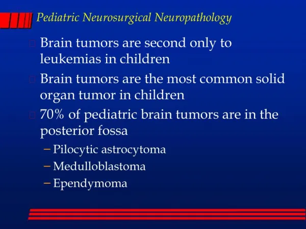
What is a brain tumor? • A group (mass) of abnormal cells that starts in the brain • There are over 120 different types of brain tumors, which makes effective treatment complicated.
Diagnosis of brain tumors • Diagnostic tools include: patient history, a brain scan, CT scan, MRI. • MRI provides a much greater contrast between the different soft tissues of the body than computed tomography (CT) does • The first MR image was published in 1973
MRI • How does it work? • The body is largely composed of water molecules. • Each water molecule has two hydrogen nuclei or protons • A powerful magnetic field causes the magnetic moments of some of these protons to align with the direction of the field. • The protons in different tissues return to their equilibrium state at different rates
MRI sequences • echo time - TE • repetition time – TR • T1 • T1-weighted scans use a gradient echo (GRE) sequence, with short Te and short Tr • This scan runs very fast allowing the → easy too collect high resolution 3D datasets. • T1-weighted scans provide good gray matter/white matter contrast. • T2 • Long Te and long Tr • Well suited to edema as they are sensitive to water content
MRI sequences (cont) • Diffusion MRI • Diffusion MRI measures the diffusion of water molecules in biological tissues. • If molecules in a particular voxel diffuse principally in one direction → the majority of the fibers in this area are going parallel to that direction. • Fluid Attenuated Inversion Recovery (FLAIR) • Inversion-recovery pulse sequence used to null signal from fluids. • Susceptibility weighted imaging (SWI) • Produces an enhanced contrast magnitude image very sensitive to venous blood, hemorrhage and iron storage. • Used to enhance the detection and diagnosis of tumors • Detects traumatic brain injuries that may not be diagnosed using other methods.
Goal • Determine: • Number of tumors • Number of abnormal cells* • Tumor margins *voxels
Previous Work • Probabilistic segmentation of brain tumors based on multi-modality magnetic resonance images. 2007 • Acqire results using 21 patients. • Test it on 22nd • 2D • 3D brain tumor segmentation in MRI using fuzzy classification, symmetry analysis and spatially constrained deformable models. 2008 • Tumor detection is performed, based on selecting asymmetric areas • Apply a segmentation method based on a combination of a deformable model and spatial relations
My approach • Combine together • Apply symmetry criteria to filter out ”not interesting” regions • Use multimodality to vote for each cell* *voxel
Data • IEEE contest organizers provided the data* • MRI sequences • Sequence name • Dimensions • Data type • Voxel to World matrix * The dataset is courtesy of Prof. B. Terwey, Klinikum Mitte, Bremen, Germany.
The process • For each sequence in the data • Convert to physical space • Register • Calculate Symmetry • Calculate Gradient
The process, step 1 • Convert from computational space to physical
The process, step 2 • Registeration • MedINRIA • Algorithm: manual landmark based • Non linear transformation
The process, step 3 • Find asymmetry : • If a[i,j] ~= a[n-i, j] a[i,j] = 0; a[n-i,j] = 0;
The process, step 4 • Calculate gradient • Data values are scalars • Scalar' = Vector • Calculate Magnitude of the vector
Process (cont) • Reference – healthy brain • Generated by using BrainWeb* - Simulated MRI Volumes for Normal Brain *McConnell Brain Imaging Centre (BIC) of the Montreal Neurological Institute, McGill University.
Process (cont) • Calculate probability • Voting function • If Healthy cell gradient < X && tumor cell gradient > Y Higher probability that the cell is abnormal • Treat noise clear skull boundaries • manual registration
Results (cont) • Voxel size 0.924mm x 1.14169mm x 2.38699mm • Total brain voxels 1,222,332 • Tumor voxels 14,878 → 0.012171816 %
Tools • Algorithm implementation C++ using VTK libraries • Viewers Paraview • Registration MedINRIA
Future Work • Make a tool • Add DTI processing • Determine spatial relationship between tumor and WM fiber tracts