Uterine Leiomyomas
430 likes | 1.45k Views
Uterine Leiomyomas. Wilfred Dang and Aliza Moledina. Objectives. 4394 : Describe the gross and histologic appearance of uterine leiomyoma and state their clinical significance .

Uterine Leiomyomas
E N D
Presentation Transcript
Uterine Leiomyomas Wilfred Dang and Aliza Moledina
Objectives • 4394: Describe the gross and histologic appearance of uterine leiomyomaand state their clinical significance. • 10670: Describe etiology, risk factors, signs and symptoms, physical examination findings, investigations and management of benignuterine neoplasia.
Case • A 40 yo G5P5 woman complains of menorrhagia of 2 years duration. She states that several years ago a doctor had told her that her uterus was enlarged. Her records indicate that 1 year ago she underwent a uterine dilatation and curettage, with the tissue showing benign pathology. She takes ibuprofen but obtains no relief of her vaginal bleeding. On examination, her blood pressure is 135/80, heart rate 80 bpm, and temperature 37.6 degrees Celcius. Abdominal examination reveals a lower abdominal midline irregular mass. On pelvic examination, the cervix is anteriorly displaced. An irregular midline mass approximately 18 weeks size seems to move in conjunction with the cervix. No adnexal masses are palpated. Her pregnancy test is negative. Her hemoglobin is 86, wbc 10 and plt 160.
Case • A 40 yo G5P5 woman complains of menorrhagia of 2 years duration. She sates that several years ago a doctor had told her that her uterus was enlarged. Her records indicate that 1 year ago she underwent a uterine dilatation and curettage, with the tissue showing benignpathology. She takes ibuprofen but obtains no relief of her vaginal bleeding. On examination, her blood pressure is 135/80, heart rate 80 bpm, and temperature 37.6 degrees Celcius. Abdominal examination reveals a lower abdominal midline irregular mass. On pelvic examination, the cervix is anteriorly displaced. An irregular midline mass approximately 18 weeks size seems to move in conjunction with the cervix. No adnexal masses are palpated. Her pregnancy test is negative.Her hemoglobin is 86, wbc 10 and plt 160.
Differential Diagnosis: Abnormal Uterine Bleeding WITH STRUCTURAL ABNORMALITY (Mass) WITHOUT STRUCTURAL ABNORMALITY
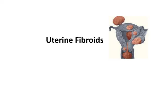
Uterine Leiomyomas (Fibroids) • Most common pelvic tumour in women • Clinically noticed in 12-25% of reproductive age women (40-50% >35 years) • Noted on pathological examination of approximately 80% of surgically excised uteri • Benign monoclonal tumours from smooth muscle cells of myometrium • Most common reason for hysterectomy in USA • Reproductive age women - typically regress after menopause
Etiology/Pathogenesis • Etiology unknown. • Estrogen as a growth promoter • Fibroids typically arise after menarche and regress after menopause • Estrogen monoclonal smooth muscle proliferation • Degenerative changes when tumour outgrows blood supply. • hyaline degeneration (most common) • cystic degeneration (from breakdown of hyaline) • red/carnerous degeneration (hemorrhage into tumour - ie pregnancy) • fatty degeneration • calcification • sarcomatous degeneration (rare) • parasitic myoma
Anatomical Classification Intramural Myomas - may distort cavity/serosal surface SubmucosalMyomas - from myometrial cells- often protude into uterine cavitySubserosalMyomas: -originate from myometrium at the serosal surface of the uterus Cervical Myomas:
Risk Factors • Race • higher in African-American women • Early menarche (< 10 years old), nulliparity • Obesity/Change in body habitus as an adult (Controversial) • Diet: beef/red meat, (Controversial), alcohol • Genetics • Hypertension • Note: smoking, increased parity found to decrease risk of fibroids
Gross Pathology • Can be single or multiple, variable in size. Note in this specimen: - uterine body is enlarged/deformed - multiple nodules, well-circumscribed, whorled appearance - uterine cavity compressed Can also have hemorrhagic areas, cystic degeneration, calcifications
Clinical Manifestations • Most asymptomatic. (60%) • Abnormal Uterine Bleeding **most common symptom** • heavy and prolonged bleeding • Inter-menstrual bleeding and post-menopausal bleeding are NOT characteristic. • Pelvic Pressure and Pain • Bulk related symptoms (ex pelvic pressure/heaviness, urinary frequency, urgency, difficulty emptying) • Constipation/Bloating (rare) • Acute Pelvic Pain • fibroid degeneration, fibroid torsion • Dysmenorrhea • Dyspareunia (deep) • Infertility/Recurrent pregnancy loss (Distortion of Uterine Cavity) • Pregnancy complications
Physical Examination • Bimanual pelvic examination • enlarged uterus • Speculum examination • prolapsed submucosal fibroid (infrequently), cervical contour
Laboratory Tests • CBC • Menorrhagia • Query Anemia?
Diagnostic Investigations Imaging • Transvaginal Ultrasound (TVS) • 95-100% sensitive • Most widely used modality • Accessible • Cost-effective • Used to determine location and size of fibroid Hypoechoicthickening around arrowheads
Diagnostic Investigations • Saline Infusion Sonography (Sonohysterography) • Improves characterization of the extent of protrusion into the endometrial cavity by submucousmyomas • Pearls: Good for differentiating endometrial polyps from submucosalmyomas • Endometrial polyps will be homogenous hyperechoic masses • Submucosalmyomas are more heterogenous and fed by multiple vessels • Masses >20mm are usually fibroids • Pearls: Good for determining the subtype of submucosalmyoma • Type 0 – Intracavitary • Type I – Less than 50% in the uterine wall • Type II – Greater than 50% in the uterine wall
Diagnostic Investigations • Magnetic Resonance Imaging • Expensive • Reserved for complex surgical planning • Can be used to differentiate fibroids from other adnexal masses • Also to distinguish fibroids from leiomyosarcomas, before uterine artery embolization (UAE) • Diagnostic Hysteroscopy • Can be performed in office • Saline or CO2 distension media = Panoramic • Without media = Contact
Diagnostic Investigations • Endometrial Biopsy • Rule out uterine cancer
Management Principles • Treatment choice based on: • Type, severity of symptoms • Size • Location • Patient age • Reproductive plans • Treat anemia if present • Treatment Options: • Conservative • Medical • Surgical
Conservative Management • Continue to Follow-up and Observe If: • Symptoms absent or minimal • Fibroids <6-8 cm or stable in size • Not submucosal • submucosalfibroids are more likely to be symptomatic • Currently pregnant due to increased risk of bleeding • follow-up U/S if symptoms progress
When to Treat • Only If: • Rapidly enlarging • Intracavitary (Submucosal Type) • Menorrhagia or menometrorrhagia • Symptomatic
Medical (Symptomatic) Management Pain • NSAIDs Hormonal Therapies (Continuous) • Estrogen–progestin contraceptives (OCP) • Levonorgestrel–releasing intrauterine system (“Mirena IUD”) • Progestin injections (Depot Medroxy Progesterone Acetate – DMPA) • Progestin pills • Selective progesterone receptor modulators • Gonadotropin-releasing hormone (GnRH) agonists, antagonists • Shrinks fibroids Antifibrinolytics (can be taken with Hormonal Therapies) • Slows bleeding quickly • Good if pregnancy is desired
Prophylactic Therapy • Prophylactic therapy to avoid potential future complications from myomas or their treatment is not recommended. • EXCEPTIONS: • Significant submucosalleiomyomas who are contemplating pregnancy • Prevent miscarriage • Myomectomy • Ureteral compression leading to moderate or severe hydronephrosis • Prevent urinary tract obstruction
Back to Our Patient • 1) Treat her anemia • Give iron supplements • 2) Check Size and Location • 3) Check desire for Pregnancy (Does she want pregnancy #6!?) • 4)) Offer Medical Management vs Surgery • Medical – Symptom Management • Myomectomy – Conserves Fertility • Other Surgeries – Affect Fertility but maybe more definite treatment (e.g. hysterectomy)
References • Uptodate • Toronto Notes • Dr. Sony Singh and Dr. Mina Wesa – “Abnormal Uterine Bleeding” Lecture Slides • Radiopedia