Download
1 / 71
730 likes | 879 Views
Herpesviruses. An Overview. Properties of herpesviruses. Enveloped double stranded DNA viruses. Genome consisits of long and short fragments which may be orientated in either direction, giving a total of 4 isomers. Three subfamilies: Alphaherpesviruses - HSV-1, HSV-2, VZV

E N D
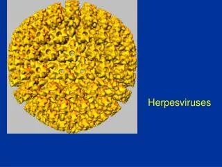
Herpesviruses An Overview
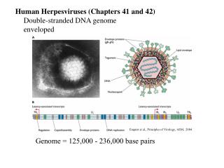
Properties of herpesviruses • Enveloped double stranded DNA viruses. • Genome consisits of long and short fragments which may be orientated in either direction, giving a total of 4 isomers. • Three subfamilies: • Alphaherpesviruses - HSV-1, HSV-2, VZV • Betaherpesviruses - CMV, HHV-6, HHV-7 • Gammaherpesviruses - EBV, HHV-8 • Set up latent or persistent infection following primary infection • Reactivation are more likely to take place during periods of immunosuppression • Both primary infection and reactivation are likely to be more serious in immunocompromised patients.
Herpesvirus Particle HSV-2 virus particle. Note that all herpesviruses have identical morphology and cannot be distinguished from each other under electron microscopy. (Linda Stannard, University of Cape Town, S.A.)
Properties • Belong to the alphaherpesvirus subfamily of herpesviruses • Double stranded DNA enveloped virus with a genome of around 150 kb • The genome of HSV-1 and HSV-2 share 50 - 70% homology. • They also share several cross-reactive epitopes with each other. There is also antigenic cross-reaction with VZV. • Man is the only natural host for HSV.
Epidemiology (1) • HSV is spread by contact, as the virus is shed in saliva, tears, genital and other secretions. • By far the most common form of infection results from a kiss given to a child or adult from a person shedding the virus. • Primary infection is usually trivial or subclinical in most individuals. It is a disease mainly of very young children ie. those below 5 years. • There are 2 peaks of incidence, the first at 0 - 5 years and the second in the late teens, when sexual activity commences. • About 10% of the population acquires HSV infection through the genital route and the risk is concentrated in young adulthood.
Epidemiology (2) • Generally HSV-1 causes infection above the belt and HSV-2 below the belt. In fact, 40% of clinical isolates from genital sores are HSV-1, and 5% of strains isolated from the facial area are HSV-2. This data is complicated by oral sexual practices. • Following primary infection, 45% of orally infected individuals and 60% of patients with genital herpes will experience recurrences. • The actual frequency of recurrences varies widely between individuals. The mean number of episodes per year is about 1.6.
Pathogenesis • During the primary infection, HSV spreads locally and a short-lived viraemia occurs, whereby the virus is disseminated in the body. Spread to the to craniospinal ganglia occurs. • The virus then establishes latency in the craniospinal ganglia. • The exact mechanism of latency is not known, it may be true latency where there is no viral replication or viral persistence where there is a low level of viral replication. • Reactivation- It is well known that many triggers can provoke a recurrence. These include physical or psychological stress, infection; especially pneumococcal and meningococcal, fever, irradiation; including sunlight, and menstruation.
Clinical Manifestations HSV is involved in a variety of clinical manifestations which includes ;- 1. Acute gingivostomatitis 2. Herpes Labialis (cold sore) 3. Ocular Herpes 4. Herpes Genitalis 5. Other forms of cutaneous herpes 7. Meningitis 8. Encephalitis 9. Neonatal herpes
Oral-facial Herpes • Acute Gingivostomatitis • Acute gingivostomatitis is the commonest manifestation of primary herpetic infection. • The patient experiences pain and bleeding of the gums. 1 - 8 mm ulcers with necrotic bases are present. Neck glands are commonly enlarged accompanied by fever. • Usually a self limiting disease which lasts around 13 days. • Herpes labialis (cold sore) • Following primary infection, 45% of orally infected individuals will experience reactivation. The actual frequency of recurrences varies widely between individuals. • Herpes labialis (cold sore) is a recurrence of oral HSV. • A prodrome of tingling, warmth or itching at the site usually heralds the recurrence. About 12 hours later, redness appears followed by papules and then vesicles.
Ocular Herpes HSV causes a broad spectrum of ocular disease, ranging from mild superficial lesions involving the external eye, to severe sight-threatening diseases of the inner eye. Diseases caused include the following:- • Primary HSV keratitis – dendritic ulcers • Recurrent HSV keratitis • HSV conjunctivitis • Iridocyclitis, chorioretinitis and cataract
Genital Herpes • Genital lesions may be primary, recurrent or initial. • Many sites can be involved which includes the penis, vagina, cervix, anus, vulva, bladder, the sacral nerve routes, the spinal and the meninges. The lesions of genital herpes are particularly prone to secondary bacterial infection eg. S.aureus, Streptococcus, Trichomonas and Candida Albicans. • Dysuria is a common complaint, in severe cases, there may be urinary retention. • Local sensory nerves may be involved leading to the development of a radiculitis. A mild meningitis may be present. • 60% of patients with genital herpes will experience recurrences. Recurrent lesions in the perianal area tend to be more numerous and persists longer than their oral HSV-1 counterparts.
Herpes Simplex Encephalitis • Herpes Simplex encephalitis is one of the most serious complications of herpes simplex disease. There are two forms: • Neonatal – there is global involvement and the brain is almost liquefied. The mortality rate approaches 100%. • Focal disease – the temporal lobe is most commonly affected. This form of the disease appears in children and adults. It is possible that many of these cases arise from reactivation of virus. The mortality rate is high (70%) without treatment. • It is of utmost importance to make a diagnosis of HSE early. It is general practice that IV acyclovir is given in all cases of suspected HSE before laboratory results are available.
Neonatal Herpes Simplex (1) • Incidence of neonatal HSV infection varies inexplicably from country to country e.g. from 1 in 4000 live births in the U.S. to 1 in 10000 live births in the UK • The baby is usually infected perinatally during passage through the birth canal. • Premature rupturing of the membranes is a well recognized risk factor. • The risk of perinatal transmission is greatest when there is a florid primary infection in the mother. • There is an appreciably smaller risk from recurrent lesions in the mother, probably because of the lower viral load and the presence of specific antibody • The baby may also be infected from other sources such as oral lesions from the mother or a herpetic whitlow in a nurse.
Neonatal Herpes Simplex (2) • The spectrum of neonatal HSV infection varies from a mild disease localized to the skin to a fatal disseminated infection. • Infection is particularly dangerous in premature infants. • Where dissemination occurs, the organs most commonly involved are the liver, adrenals and the brain. • Where the brain is involved, the prognosis is particularly severe. The encephalitis is global and of such severity that the brain may be liquefied. • A large proportion of survivors of neonatal HSV infection have residual disabilities. • Acyclovir should be promptly given in all suspected cases of neonatal HSV infection. • The only means of prevention is to offer caesarean section to mothers with florid genital HSV lesions.
Other Manifestations • Disseminated herpes simplex are much more likely to occur in immunocompromised individuals. The widespread vesicular resembles that of chickenpox. Many organs other than the skin may be involved e.g. liver, spleen, lungs, and CNS. • Other cutaneous manifestations include • eczema herpeticum which is potentially a serious disease that occurs in patients with eczema. • Herpetic whitlow which arise from implantation of the virus into the skin and typically affect the fingers. • “zosteriform herpes simplex". This is a rare presentation of herpes simplex where HSV lesions appear in a dermatomal distribution similar to herpes zoster.
Laboratory Diagnosis • Direct Detection • Electron microscopy of vesicle fluid - rapid result but cannot distinguish between HSV and VZV • Immunofluorescence of skin scrappings - can distinguish between HSV and VZV • PCR - now used routinely for the diagnosis of herpes simple encephalitis • Virus Isolation • HSV-1 and HSV-2 are among the easiest viruses to cultivate. It usually takes only 1 - 5 days for a result to be available. • Serology • Not that useful in the acute phase because it takes 1-2 weeks for before antibodies appear after infection. Used to document to recent infection.
Cytopathic Effect of HSV in cell culture: Note the ballooning of cells. (Linda Stannard, University of Cape Town, S.A.) Positive immunofluorescence test for HSV antigen in epithelial cell. (Virology Laboratory, New-Yale Haven Hospital)
Management At present, there are only a few indications of antiviral chemotherapy, with the high cost of antiviral drugs being a main consideration. Generally, antiviral chemotherapy is indicated where the primary infection is especially severe, where there is dissemination, where sight is threatened, and herpes simplex encephalitis. Acyclovir – this the drug of choice for most situations at present. It is available in a number of formulations:- • I.V. (HSV infection in normal and immunocompromised patients) • Oral (treatment and long term suppression of mucocutaneous herpes and prophylaxis of HSV in immunocompromised patients) • Cream (HSV infection of the skin and mucous membranes) • Ophthalmic ointment Famciclovir and valacyclovir – oral only, more expensive than acyclovir. Other older agents – e.g. idoxuridine, trifluorothymidine, Vidarabine (ara-A). • These agents are highly toxic and is suitable for topical use for opthalmic infection only
Properties • Belong to the alphaherpesvirus subfamily of herpesviruses • Double stranded DNA enveloped virus • Genome size 125 kbp, long and short fragments with a total of 4 isometric forms. • One antigenic serotype only, although there is some cross reaction with HSV.
Epidemiology • Primary varicella is an endemic disease. Varicella is one of the classic diseases of childhood, with the highest prevalence occurring in the 4 - 10 years old age group. • Varicella is highly communicable, with an attack rate of 90% in close contacts. • Most people become infected before adulthood but 10% of young adults remain susceptible. • Herpes zoster, in contrast, occurs sporadically and evenly throughout the year.
Pathogenesis • The virus is thought to gain entry via the respiratory tract and spreads shortly after to the lymphoid system. • After an incubation period of 14 days, the virus arrives at its main target organ, the skin. • Following the primary infection, the virus remains latent in the cerebral or posterior root ganglia. In 10 - 20% of individuals, a single recurrent infection occurs after several decades. • The virus reactivates in the ganglion and tracks down the sensory nerve to the area of the skin innervated by the nerve, producing a varicellaform rash in the distribution of a dermatome.
Varicella • Primary infection results in varicella (chickenpox) • Incubation period of 14-21 days • Presents fever, lymphadadenopathy. a widespread vesicular rash. • The features are so characteristic that a diagnosis can usually be made on clinical grounds alone. • Complications are rare but occurs more frequently and with greater severity in adults and immunocompromised patients. • Most common complication is secondary bacterial infection of the vesicles. • Severe complications which may be life threatening include viral pneumonia, encephalititis, and haemorrhagic chickenpox.
Herpes Zoster (Shingles) • Herpes Zoster mainly affect a single dermatome of the skin. • It may occur at any age but the vast majority of patients are more than 50 years of age. • The latent virus reactivates in a sensory ganglion and tracks down the sensory nerve to the appropriate segment. • There is a characteristic eruption of vesicles in the dermatome which is often accompanied by intensive pain which may last for months (postherpetic neuralgia) • Herpes zoster affecting the eye and face may pose great problems. • As with varicella, herpes zoster in a far greater problem in immunocompromised patients in whom the reactivation occurs earlier in life and multiple attacks occur as well as complications. • Complications are rare and include encephalitis and disseminated herpes zoster.
Congenital VZV Infection • 90% of pregnant women already immune, therefore primary infection is rare during pregnancy. • Primary infection during pregnancy carries a greater risk of severe disease, in particular pneumonia. First 20 weeks of Pregnancy • Up to 3% chance of transmission to the fetus, recognised congenital varicella syndrome; • Scarring of skin • Hypoplasia of limbs • CNS and eye defects • Death in infancy normal
Neonatal Varicella • VZV can cross the placenta in the late stages of pregnancy to infect the fetus congenitally. • Neonatal varicella may vary from a mild disease to a fatal disseminated infection. • If rash in mother occurs more than 1 week before delivery, then sufficient immunity would have been transferred to the fetus. • Zoster immunoglobulin should be given to susceptible pregnant women who had contact with suspected cases of varicella. • Zoster immunoglobulin should also be given to infants whose mothers develop varicella during the last 7 days of pregnancy or the first 14 days after delivery.
Laboratory Diagnosis The clinical presentations of varicella or zoster are so characteristic that laboratory confirmation is rarely required. Laboratory diagnosis is required only for atypical presentations, particularly in the immunocompromised. • Virus Isolation - rarely carried out as it requires 2-3 weeks for a results. • Direct detection - electron microscopy may be used for vesicle fluids but cannot distinguish between HSV and VZV. Immunofluorescense on skin scrappings can distinguish between the two. • Serology - the presence of VZV IgG is indicative of past infection and immunity. The presence of IgM is indicative of recent primary infection.
Cytopathic Effect of VZV Cytopathic Effect of VZV in cell culture: Note the ballooning of cells. (Coutesy of Linda Stannard, University of Cape Town, S.A.)
Management • Uncomplicated varicella is a self limited disease and requires no specific treatment. However, acyclovir had been shown to accelerate the resolution of the disease and is prescribed by some doctors. • Acyclovir should be given promptly immunocompromised individuals with varicella infection and normal individuals with serious complications such as pneumonia and encephalitis. • herpes zoster in a healthy individual is not normally a cause for concern. The main problem is the management of the postherpetic neuralgia. • The International Herpes Management Forum recommends that antiviral therapy should be offered routinely to all patients over 50 years of age presenting with herpes zoster. • Three drugs can be used for the treatment of herpes zoster: acyclovir, valicyclovir, and famciclovir. There appears to be little difference in efficacy between them.
Prevention • Preventive measures should be considered for individuals at risk of contracting severe varicella infection e.g. leukaemic children, neonates, and pregnant women • Where urgent protection is needed, passive immunization should be given. Zoster immunoglobulin (ZIG) is the preparation of choice but it is very expensive. Where ZIG is not available, HNIG should be given instead. • A live attenuated vaccine is available. There had been great reluctance to use it in the past, especially in immunocompromised individuals since the vaccine virus can become latent and reactivate later on. • However, recent data suggests that the vaccine is safe, even in children with leukaemia provided that they are in remission. • It is highly debatable whether universal vaccination should be offered since chickenpox and shingles are normally mild diseases.
Properties • Belong to the betaherpesvirus subfamily of herpesviruses • double stranded DNA enveloped virus • Nucleocapsid 105nm in diameter, 162 capsomers • The structure of the genome of CMV is similar to other herpesviruses, consisting of long and short segments which may be orientated in either direction, giving a total of 4 isomers. • A large no. of proteins are encoded for, the precise number is unknown.
Epidemiology • CMV is one of the most successful human pathogens, it can be transmitted vertically or horizontally usually with little effect on the host. • Transmission may occur in utero, perinatally or postnatally. Once infected, the person carries the virus for life which may be activated from time to time, during which infectious virions appear in the urine and the saliva. • Reactivation can also lead to vertical transmission. It is also possible for people who have experienced primary infection to be reinfected with another or the same strain of CMV, this reinfection does not differ clinically from reactivation. • In developed countries with a high standard of hygiene, 40% of adolescents are infected and ultimately 70% of the population is infected. In developing countries, over 90% of people are ultimately infected.
Pathogenesis • Once infected, the virus remains in the person for life and my be reactivated from time to time, especially in immunocompromised individuals. • The virus may be transmitted in utero, perinatally, or postnatally. Perinatal transmission occurs. • Perinatal infection is acquired mainly through infected genital secretions, or breast milk. Overall, 2 - 10% of infants are infected by the age of 6 months worldwide. Perinatal infection is thought to be 10 times more common than congenital infection. • Postnatal infection mainly occurs through saliva. Sexual transmission may occur as well as through blood and blood products and transplanted organ.
Clinical Manifestations • Congenital infection - may result in cytomegalic inclusion disease • Perinatal infection - usually asymptomatic • Postnatal infection - usually asymptomatic. However, in a minority of cases, the syndrome of infectious mononucleosis may develop which consists of fever, lymphadenopathy, and splenomegaly. The heterophil antibody test is negative although atypical lymphocytes may be found in the blood. • Immunocompromised patients such as transplant recipients and AIDS patients are prone to severe CMV disease such as pneumonitis, retinitis, colitis, and encephalopathy. • Reactivation or reinfection with CMV is usually asymptomatic except in immunocompromised patients.
Congenital Infection • Defined as the isolation of CMV from the saliva or urine within 3 weeks of birth. • Commonest congenital viral infection, affects 0.3 - 1% of all live births. The second most common cause of mental handicap after Down's syndrome and is responsible for more cases of congenital damage than rubella. • Transmission to the fetus may occur following primary or recurrent CMV infection. 40% chance of transmission to the fetus following a primary infection. • May be transmitted to the fetus during all stages of pregnancy. • No evidence of teratogenecity, damage to the fetus results from destruction of target cells once they are formed.
Cytomegalic Inclusion Disease • CNS abnormalities - microcephaly, mental retardation, spasticity, epilepsy, periventricular calcification. • Eye - choroidoretinitis and optic atrophy • Ear - sensorineural deafness • Liver - hepatosplenomegaly and jaundice which is due to hepatitis. • Lung - pneumonitis • Heart - myocarditis • Thrombocytopenic purpura, Haemolytic anaemia • Late sequelae in individuals asymptomatic at birth - hearing defects and reduced intelligence.
Laboratory Diagnosis (1) • Direct detection • biopsy specimens may be examined histologically for CMV inclusion antibodies or for the presence of CMV antigens. However, the sensitivity may be low. • The pp65 CMV antigenaemia test is now routinely used for the rapid diagnosis of CMV infection in immunocompromised patients. • PCR for CMV-DNA is used in some centers but there may be problems with interpretation.
CMV pp65 antigenaemia test (Virology Laboratory, New-Yale Haven Hospital)
Laboratory Diagnosis (2) • Virus Isolation • conventional cell culture is regarded as gold standard but requires up to 4 weeks for result. • More useful are rapid culture methods such as the DEAFF test which can provide a result in 24-48 hours. • Serology • the presence of CMV IgG antibody indicates past infection. • The detection of IgM is indicative of primary infection although it may also be found in immunocompromised patients with reactivation.
Cytopathic Effect of CMV (Courtesy of Linda Stannard, University of Cape Town, S.A.)
DEAFF test for CMV (Virology Laboratory, New-Yale Haven Hospital)
Treatment • Congenital infections - it is not usually possible to detect congenital infection unless the mother has symptoms of primary infection. If so, then the mother should be told of the chances of her baby having cytomegalic inclusion disease and perhaps offered the choice of an abortion. • Perinatal and postnatal infection - it is usually not necessary to treat such patients. • Immunocompromised patients - it is necessary to make a diagnosis of CMV infection early and give prompt antiviral therapy. Anti-CMV agents in current use are ganciclovir, forscarnet, and cidofovir.
Prevention • No licensed vaccine is available. There is a candidate live attenuated vaccine known as the Towne strain but there are concerns about administering a live vaccine which could become latent and reactivates. • Prevention of CMV disease in transplant recipients is a very complicated subject and varies from center to center. It may include the following measures. • Screening and matching the CMV status of the donor and recipient • Use of CMV negative blood for transfusions • Administration of CMV immunoglobulin to seronegative recipients prior to transplant • Give antiviral agents such as acyclovir and ganciclovir prophylactically.