Download
1 / 178
1.84k likes | 1.95k Views
Maternal Physiological Changes during Pregnancy , Labor and Puerperium. ASSOCIATE PROFESSOR IOLANDA BLIDARU MD, PhD. Pregnancy is a period of adaptation for :. The needs of the fetus Meeting the stress of pregnancy and labour. THE GENITAL CHANGES. (A) The whole uterus. 1 - Size.

E N D
Maternal Physiological Changes during Pregnancy, Labor and Puerperium ASSOCIATE PROFESSOR IOLANDA BLIDARU MD, PhD
Pregnancy is a period of adaptation for : • The needs of the fetus • Meeting the stress of pregnancy and labour
1 - Size increase from 7.5 x 5 x 2.5 cm in nonpregnant states to 36 x 25 x 20 cm at term i.e. the volume increase 1000 time
increases from 50 gm in nonpregnant state to 1000 gm at term 2 - Weight
pyriform in the nonpregnant state , becomes globular at 8th week, then ovoid by 16th week till term . 3 -Shape
4 - Position with ascent from the pelvis , the uterus usually undergoes rotation with tilting to the right (dextrorotation) due to the presence of the rectosegmoid colon on the left side.
5 - Consistency : becomes progressively softer due to : 1 - Increased vascularity 2 - Presence of amniotic fluid
6 -Contractility from the first trimester onwards , the uterus undergoes irregular painless contractions (Braxton Hicks contractions) . They may cause some discomfort late in pregnancy and may account for false labour pain .
7- Capacity increases from 4 ml in non-pregnant state to 4-5000 ml at term
1 - Hypertrophy (estrogen effect) + hyperplasia (progesterone effect)2 - the fetus exerts a direct stretch
2 - Formation of the lower uterine segment (L.U.S.) from the isthmus
Formation of lower uterine segment After 12 weeks, the isthmus(0.5cm) starts to expand gradually to form the lower uterine segment which measures 10-11 cm in length at term
Upper Uterine Segment • Peritoneum: Firmly-attached • Myometrium:3 layers; outer longitudinal, middle oblique (interlacing network) and inner circular. • The middle layer forms 8-shaped fibers around the blood vessels to control postpartum hemorrhage
Upper Uterine Segment • Decidua:Well-developed • Membranes:Firmly-attached • Activity:Active, contracts, retracts and becomes thicker during labour.
Lower Uterine Segment • Peritoneum: Loosely-attached • Myometrium : 2 layers; outer longitudinal and inner circular.
Lower Uterine Segment • Decidua: Poorly-developed • Membranes: Loosely- attached. • Activity:Passive, dilates, stretches and becomes thinner during labour
The junction between the upper uterine segment (U.U.S.) which is thick and the lower uterine segment which is thin is called the physiologic contraction ringat the level of the symphysis pubis (not seen or felt)
1 - Uterine artery lumen: is doubled and its blood flow increases 5 times 2 - Myometrial and decidual arteries (spiral arteries) undergo fibrinoid degeneration due to 2 waves of trophoblastic migration , so they become dilated to be the uteroplacentalarteries
Uterine blood flow increases progressively and reaches about 500 ml / minute at term
(D) Changes in the cervix : 1 - It becomes hypertrophied , soft and bluish in colour due to oedema and increased vascularity.
2 - Soon after conception, a thick cervical secretion obstructs the cervical canal forming a mucous plug . 3 - The endocervical epithelium proliferates and / or everts forming cervical ectopy(previously called erosion) (sometimes).
4 - There are 3 principal structural components of the cervix: smooth muscle (only 10%), collagen and connective tissue or extracellular matrix (ground substance).
5 -The cervical ripening process- changes in collagen (breakdown and rearrangement of the fibers), connective tissue and its ground substance (alterations of the various glycosaminoglycans). 6 - In the ripening of uterine cervix there are involved: PG E2 and PG F2 alpha,E, P and relaxin.
(E) Changes in fallopian tubes and round & broad ligaments : Inactive , elongated , marked increase in vascularity There may be broad ligament varicose veins
(F) Changes in the vagina : The vagina becomes soft , warm , moist with increased secretion and violet in colour (Chadwick's sign) due to increased vascularity
(G) Changes in the vulva : • It becomes soft, violet in colour • Oedema and varicosities may develop
(H) Changes in the ovaries 1 - Both ovaries are enlarged due to increased vascularity and oedema particularly the ovary which contains the corpus luteum .
(H) Changes in the ovaries 2 - Corpus luteum continues to grow till 7 - 8 weeks, then it stops growing. It becomes inactive and starts degeneration at 12 weeks (degeneration is completed after labour)
Corpus luteum secretes 1.estrogen 2.progesterone 3.relaxin hormones
(H) Changes in the ovaries 3 - Ovulation ceases during pregnancy due to pituitary inhibition by the high levels of oestrogen and progesterone
Relaxin is a protein hormone. • Its complete role in pregnancy is debated. • It may induce softness and effacement of the cervix.
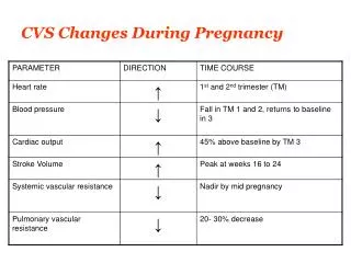
(A) Blood volume The total blood volume increases steadily from early pregnancy to reach a maximum of 35-45 % above the non-pregnant level at 32 week .
Plasma volume : Increases from 2600 ml by ± 45 % (1200 in the 1st pregnancy) and 1500 ml in subsequent pregnancies After delivery, it decreases rapidly by 600-800ml.
Red blood cell mass : • Increases from 1400 ml (nonpregnant) by 30 % (± 450 ml) due to increased production resulting from erythropoietin action • The increase is steady till full term.
The increase in plasma volume is more than the increase in red blood cell mass (Hb mass) resulting in haemodilution (physiologic anemia)
However, the minimal Hb. accepted is 10-11 gm%
Values of increased blood volume 1 - Meets increased demands for uterus, baby .... etc. 2 - Protects against supine hypotension syndrome. 3 - Protects against fluid loss at delivery.
Increased blood volume more than the increase in red cell mass, leads to decreased blood viscosity which leads to adecrease in peripheral resistance
1 - Decreased Hb % and RBCs % : • Erythrocytes decrease from 4.5 million / mm3 to 3.7 million / mm3 (due to the relative increase in plasma volume more than red cell mass).