Download
1 / 33
330 likes | 405 Views
Novel Tools for (Functional) Magnetic Resonance Image Analysis Development and Implementation in the Scientific and Statistical Computing Core Robert W Cox and a cast of several. MR-scanner. Raw data. Scanner Subject Stimulus Delivery. Reconstruction Distortion correction. BOLD EPI.

E N D
Novel Tools for(Functional)Magnetic Resonance Image AnalysisDevelopment and Implementation in the Scientific and Statistical Computing CoreRobert W Coxand a cast of several
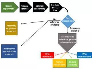
MR-scanner Raw data Scanner Subject Stimulus Delivery Reconstruction Distortion correction BOLD EPI Experiment design Co-registration Anatomy Function BOLD signal Statistical models Inference Func. & Anat. Group analysis
} Rich source of ideas for novel tools Scientific & Statistical Computing Core • Develop and implement new methodologies to meet user needs • Consult with IRP users/groups regarding • Experimental design • Processing methods and tools • Statistical inferences • Conduct classes on designing and processing FMRI experiments • Answer FMRI/MRI questions on message board • Distribute & maintain our open-source software tools • Facilitate cross-talk between different FMRI tools: • AFNI, FSL, FMRIstat, FreeSurfer, Caret, SPM, …
AFNI + SUMA • AFNI= collection of programs for FMRI analysis • Visualization • 2D, 3D, time-series, cortical surface (SUMA) • Time Series Analysis • Linear & nonlinear regression • Statistics on 3D Image Collections • 1-5 way ANOVA; non-parametrics; SEM • Data editing tools • Spatial and temporal filtering • 3D image registration • Clustering; ROI drawing & Atlas-based ROIs
The AFNI / SSCC Philosophy • Enable users to stay close to their data • Save intermediate results • Look at images and data in connected ways • User controls processing steps and parameters • Everyone has an opinion • Special problems need special solutions • Efficient (fast) implementations • Things that are easy and fast to do will get done more often • Help the users • Until our patience runs out
Next Set of Slides Features Added to AFNI and SUMA in Response to User Requests and/or Problems/Complaints (at least in part)
Feature: Atlases • Problem: Navigating in a complicated folded up 3D object (i.e., the brain) with few easily recognized landmarks • Solution: Coordinate-based brain atlases • Accepting the 5-10 mm uncertainty of brain coordinates • Atlas #1: Talairach-Tournoux atlas • As parsed by Peter Fox’s group at UT San Antonio • Atlas #2: Cytoarchitectonic atlases from Karl Zilles’ group at Forschungszentrum Jülich • 10 brains being sliced & diced & stained & scanned • About 40% complete at this time • Where Am I? + Jump To + Colorization + ROIs • Plans: keep up with Zilles; Animal atlases? …
A Feature: Skull Stripping • Problem: other skull stripping software (e.g., BET in FSL) didn’t work reliably enough • Solution was to re-visit problem from scratch, and build on BET’s surface growing algorithm • Then add new features: special knowledge about where the eyes are likely to be; 3D edges; etc. • Then test it on the hard cases from NIH (ab)users • Extra feature: extend it to monkey images • Plans: continue testing and improvements
A Feature: De-Spiking • Problem: occasional big spikes in echo planar images gathered for functional MRI • Problem eventually traced to gradient coil • In the meantime: can studies be saved? • Wrecks the standard time series analysis
Feature: Amplitude Modulated FMRI • Situation: Each stimulus event comes with an auxiliary parameter • May be measured (GSR, reaction time, …) or may be determined by experimenter • Want to determine if FMRI response magnitude is proportional to this auxiliary parameter • Solution was to add amplitude modulated regressors to AFNI’s 3dDeconvolve program • Two regressors per condition • First is: each stimulus response identical • Second is: each stimulus response proportional to auxiliary parameter for that stimulus • Plans: 2-3 params/event; event-wise amplitudes
Feature: Nonlinear Regression Models • Pharmacological models for time series analysis • AFNI’s nonlinear regression program 3dNLfim • Michaelis-Menton dynamics for BOLD FMRI with psychoactive drugs (e.g., ethanol) • Dynamic Contrast Enhanced MRI for quantifying Gd contrast leakage through blood-brain barrier
Feature: Smart Blurring • FMRI time series datasets are often smoothed (blurred) in space to • Reduce noise (by averaging) • Increase intra-subject activation “blob” overlap • Blurring brain & non-brain signals together is silly • When combining data from different scanners (i.e., multi-center studies), image smoothness varies • Should blur images until they have the same level of smoothness so that inter-scanner combinations make statistical sense • Developed a method for blurring inside a mask that stops when image noise reaches specified level of smoothness:
Feature: Structural Equation Modeling • SEM is a form of connectivity analysis • Input: correlations between activated ROIs • Regions where the activations fluctuate in strength together will be more highly correlated • Input: connectivity diagram between ROIs • Output: strength of connections • Can also search for “better” fitting connections
Feature: All-in-One Analysis Program • Common complaint: “AFNI is tooooooo hard to use” • Analysis of single subject data involves several steps, each instantiated in separate programs • Registration, smoothing, normalizing, model analysis • Solution is a program afni_proc.py that will run all these programs in a coherent sequence • Intermediate results are saved to make it possible to track backwards when results are confusing • This script is not intended to let the user avoid understanding the data analysis process!
Feature: Diffusion Tensor Analysis • Goal: Compute the Diffusion Tensor (etc.) from Diffusion Weighted image collections • Problem #1: log+linear method is inaccurate in highly anisotropic locations (the cool places to be) • Problem #2: published nonlinear solution methods not available in open-source software • Solution was to create and implement an efficient robust nonlinear method for finding the diffusion tensor D in each voxel • Also, a optional nonlinear image smoother (2D and 3D) to reduce noise in homogenous areas • Our code now incorporated into DTI Query, an open-source tractography program from Stanford
Feature: Inter-Modality Registration • Goal: Efficiently align 3D volumes acquired with different imaging contrasts • Solution is a general program using histogram-based measurements of image matching (e.g., mutual information) • This one is still very much a work-in-progress • Works pretty well on “simple” cases (e.g., whole-brain to whole-brain) • Dealing with partial-brain to whole-brain and with brain images that have holes in them is less reliable right now • Also want to add non-affine warping capabilities
A Example: Inter-Modality Registration Skull Stripped MRI … masked CT … CT overlaid on MRI in color - unaligned … CT overlaid on MRI in color - aligned
Feature: Analysis of Mn Contrast MRI • Mn is an MRI contrast agent and a calcium analog • Goal: time-dependent in vivo tract tracing in monkeys • Problems abound: • Like FMRI, signal changes are small • Other artifacts from day-to-day scanning are larger • Simple image subtraction isn’t reliable • Next 3 slides: some data and results …
A Mn Data: Different Days
A Mn Data: Subtract & t-Test
A Mn Data: Cleverer t-Test
Next Set of Slides Features Added to AFNI and SUMA in Response to Our Own Crazy Thoughts (mostly)
Realtime FMRI Functional activation& Motion estimation in realtime AFNI Dimon Feedback Receiver MR Scanner, Image Files
Movie capture from SUMA • Activation map projected from AFNI Surface-Based Analyses • Create cortical surface models, project 3D data to these surfaces, analyze in that space • Respects geometry and topology of cortex • Most AFNI statistical tools now work with image data defined over surfaces as well as over 3D volumes
NIfTINeuroimaging Informatics Technology Initiative • Goal: facilitate inter-operability of FMRI data analysis software • First fruit: NIfTI-1.1 standard for storing datasets defined over 3D volumes (plus time axis) • Works with AFNI, FSL, SPM, BrainVoyager, … • Agreement is not a one-time thing • Ongoing process is needed to deal with compatibility, extensions, new ideas along the same line, … • Efforts underway: • NIfTI-G: standard for storing cortical surface models (and associated data) • NIfTI-W: standard for storing non-affine spatial warps
Closely Linked Communication • Programs “talk” to each other (esp. AFNI & SUMA) • Exchange data • Issue commands - you can script many parts of the AFNI & SUMA graphical interfaces AFNI SUMA 3dSkullStrip
Developer-friendliness Realtime physiological monitoring using AFNI: Jerzy Bodurka, FIM/LBC/NIMH
L R Brain State Classification • Train Support Vector Machine (SVM) classifier on a collection of pre-categorized 3D brain images • e.g., “looking at house” and “looking at face” • Classifies new 3D images into the categories From Stephen LaConte; Emory, transitioning to Rice
Penultimate Slide • Much of our most fruitful and satisfying work comes from close and ongoing interactions with investigators that have interesting problems • Derived from studies that are pushing the envelope of deriving information from MRI • We are here to provide solutions to problems (of image analysis) • Your current short-term problems (lots of these!) • Your actual longer-term problems • What we think your future needs will be
Ultimate Slide The Team