Download
1 / 38
410 likes | 852 Views
Attention : ** Early unicellular forms evolved into multicellular forms. ** Such need to multicellularity is based on: The limited cellular diffusion and transport of nutrients and metabolites.

E N D
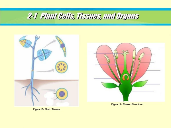
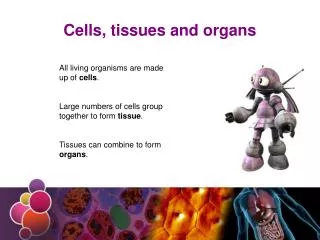
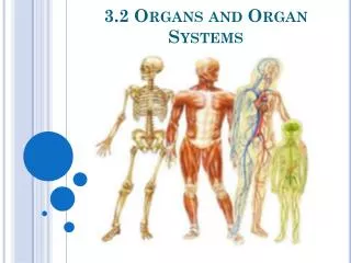
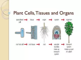
Attention: **Early unicellular forms evolved into multicellular forms.**Such need to multicellularity is based on:The limited cellular diffusion and transport of nutrients and metabolites. The result was a maximum limit on cell size followed by many cells working in coorporation with each other, which in due course, led to specialization of labor and to specialization of cells. Levels Of Organization:Cells----- Tissues----- Organs----- Organ Systems---- Organism Although cells are the ultimate building blocks of the body, these cells are organized into a higher levels of complexity in the formation of tissues.
The four basic tissue types. Epithelial tissue connective tissue (support cells, immune cells, blood cells),.muscle tissue (contractile cells), and nervous tissue. Organs represent various combinations of these four basic tissue types, which thus comprise the entire body. Each tissue type retains its fundamental character wherever it occurs.
Epithelial tissue The boundary between you and your environment is marked by a continuous surface, or epithelium, of contiguous cells.
Several features characterize epithelial tissue and distinguish it from connective tissue, muscle tissue, and nervous tissue • Epithelial tissues are arranged in sheets or layers covering the surfaces or lining the cavities of the body. • Epithelial cells are attached to one another. Special devices (intercellular junctions, tonofilaments) provide for structural integrity of the epithelium. • Their cells lack protoplasmic processes except for microvilli. • Intercellular substance is little and intercellular spaces are small.
Epithelial tissue lacks a vascular supply • Derived from all three of the embryonic germ layers. • Skin and body opinings ------- ectoderm • Lining of G.T., respiratoty system,and the glands of the G.T.--------- endoderm • Lining od kidney blood vessels --------mesoderm. • Epithelial cells are polarized. • Epithelial cells are separated from the underlying tissue by a basement membrane
The basement membrane • is a thin sheet of collagen and glycoproteins produced: • in part by the epithelial cells themselves and • in part by underlying connective tissue cells (specifically, fibroblasts). • The basement membrane serves to regulate cell behavior and can limit the spread of some neoplasms.
By Electron microscopy, basement membrane is composed of: • Amorphous Basal lamina(50 t0 100 nm thick): is a product of epithelial . It is separated from the basal cell membrane by a lucent region of 50 nm • A Reticular lamina containing reicular and collagenous fibers. • The basal lamina is seen no only in epithelial tissues but also in smooth, skeletal and cardiac muscle, in addition to fat cells.
Staining of the basement membrane • Stains deep black in silver preparations because of the reticular fibers • Stains red in PAS preparations because of the polysaccharides.
In H&E preparations: • It may appear thick such as in trachea, because of the fixed reticular lamina. • It is very thin beneathtransitional epithelium and cannot be observed by light microscope. • Basement membranes are at best inconspicuous in glomeruli of the kidney.
lumen Brush border Basement membrane The nucleus of the cell Basal side of the cells Apical side of the cells lumen basement membrane, kidney (PAS)
Classification of the epithelium I- According to the structure (number of layers of cells rest on the basement membrane): • Simple epithelium: one layer of cells • Pseudostratified epithelium: one layer of cells • Stratified epithelium: two or more layers of cells
According the shape of cells on the top: • Simple epithelium: • Simple Squamous Epithelium • SimpleCuboidal Epithelium • Simple Columnar Epithelium • Stratified epithelium: • Stratified Squamous Epithelium • Stratified cuboidal Epithelium • Stratified columnar Epithelium • Transitional Epithelium
Simple epithelium • Simple Squamous Epithelium • SimpleCuboidal Epithelium • Simple Columnar Epithelium
1- Simple squamous epithelium • Squamous: from squama, scale • consists of a single, very thin layer of flattened (squamous) cells. • Cells are large with clear or granular cytoplasm and round, oval or eccentric nuclei. • In cross section, the cytoplasm is barely visible
Simple squamous epithelium,cheekcells, top veiw. These cells are large, but quite thin, and have a prominent, protruding nucleus. A good analogy to their shape is the sunny-side-up fried egg.
Squamous epithelium Simple squamous epithelium is found inbarriers where diffusion or filtration is the basic requirement. 1- makes up the lung alveoli. The cells are very thin, side view.
2- at sites where very little activity is occuring, such as Bowman's capsule in the kidney. • Simple squamous epitheliumLateral view.
Simple epithelium Nucleus of the squamous cell Squamous cell Lateral view, simple squamous epithelium, kidney(H&E)
simple squamous Endothelium:*is a simple squamous tissue lines all blood and lymphatic vessels.**Endothelial cells may be phagocytic and can form stellate –shaped connective tissue fiberoblasts by cell division Cross section in blood vessel, lateral view of endothelium. The cells are very thin
endothelium Endothelial cell Lumen of the vessel Endothelial cells Endothelium, (H&E)
2- Simple cuboidal epithelium • consists of boxy (cuboidal) cells on the surface. • The cells are smaller and more regular than those of squamous epithelium. • In vertical section, the cells are square and contain a rounded nucleus • Locations: lining thyroid follicles,kidney tubules, salivary ducts, pancreatic ducts.
Cuboidal Epithelium, ThyroidThyroid is an endocrine gland in which simple cuboidal epithelium surrounds masses of storage protein.
Simple cuboidal and squamous epithelia in kidney (H&E), T.S.
collecting ducts collecting ducts ascending limb of the loop of Henle. Simple Cuboidal epithelium:1- Cuboidal epithelial cells with eosinophilic (i.e., pink) cytoplasm belong to the ascending limb of the loop of Henle. • 2- Cuboidal epithelial cells with relatively unstained (i.e, pale) cytoplasm belong to collecting ducts, which are larger than the tubules of the loop of Henle. (t.s in medulla)
Simple cuboidal epithelium lines the proximal and distal convoluted tubules in the kidney, t.s. in cortex.
Comparison of Duct and Blood Vessels:Simple Cuboidal epithelium is commonly encountered in glandular ducts. Small ducts typically have a simple cuboidal epithelium. Larger ducts may have a stratified cuboidal epithelium.
3- Simple columnar epithelium • In surface veiw, it appears like cuboidal; • in sections, the cells are taller than board and are rectangular, with the long axis perependicular to the free surface • The nucleus is oval and located close to the base , unless the cells are very compressed. • The free or apical portion may have a plasma membrane covered with mucous, cilia or microvilli. • lines the digestive tract and the female reproductive tract (as well as numerous other surfaces).
Types of Simple columnar epithelium • Without goblet cells: in gall bladder, stomach, • With goblet cells : intestine and respiratory tracts.
simple columnar epithelium without goblet cells. This picture is from a gall bladder. The red arrow is pointing to red blood cells (erythrocytes).
Simple columnar epithelium(without goblet cells) that form the surface of the stomach mucosa.
Gastric pit Mucous neck cells Branched gland Surface mucous cells Lamina propria Praital cells Lamina propria fundic stomach (H&E) Lamina propria Lamina propria
Goblet cell The simple columnar epithelium with goblet cellsThe villi of the small intestine are lined by a single layer of columnar cells (A). Note these cells are not as wide as they are tall with the darkly stained nuclei (B)located at the base of the cells.The cell membranes (C) are very thin but easily identified. columnar cells
Simple columnar epitheliumThe epithelial tissue layer of the small intestine contains absorptive cells, which take in nutrients, and goblet cells, which secrete mucus. The epithelial surface forms many small projections, called villi, one of which occupies most of this image.
Simple columnar with goblet cells, of villus, Small Intestine
3-Simple columnar epithelium with cilia • In which the free border is supplied with cilia e.g. Fallopian tube, vasa efferentia, ependyma,
Ependymal cells:line the ventricles of the brain and the central canal of the spinal cord are lined with The cells are often cilated and form a simple cuboidal or low columnar epithelium. The lack of tight junctions between ependymal cells allows a free exchange between cerebrospinal fluid and nervous tissue.