Download
1 / 32
340 likes | 561 Views
20. Medical physics techniques. BTEC level 3. Aim.

E N D
20. Medical physics techniques BTEC level 3
Aim • The aim of this unit is to enable learners to develop, through a practical vocational skills approach, an understanding of the important fundamental physics concepts behind medical physics techniques such as x-rays, ultrasounds, diagnostic imaging and magnetic resonance imaging (MRI) and radiotherapy. • Learners will also understand the importance of radiation safety.
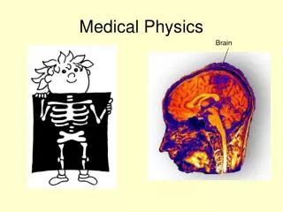
Diagnostic medicine has come a long way since the time when the best diagnosis occurred during the postmortem examination. Surgery today is faster, less invasive and more effective than ever – thanks in part to improvements in medical imaging technology. Imaging gives the doctor a clearer understanding of the patient’s condition so treatment can be planned more effectively and therapy delivered more precisely.
Nuclear medicine is providing hope for the cure of the most serious diseases, especially cancer. Radioactive materials are used in this rapidly developing branch of medicine. At the cutting edge of developments in nuclear medicine is the precise targeting needed to get the radiation to the exact site of the cancer.
Future prospects are even more exciting. Medical imaging is extending human vision into the very nature of • disease; at the cellular level it will permit diagnosis before symptoms even appear. Surgery in the future will be bloodless, painless and non-invasive. It will be powered by medical imaging systems that focus on the disease and use energy to destroy the target but preserve healthy tissue. Researchers are testing the use of highintensity ultrasound to destroy tumours identified and targeted while the patient lies in an MRI scanner.
This unit introduces learners to some of the established practices in medical physics imaging. It aims to deliver the underpinning knowledge of several of the fundamental techniques and provide a basic introduction to the more complicated theory of magnetic resonance imaging.
Learning outcomes • Know atomic structure and the physical principles of ionising radiation and ultrasound • Understand how radiopharmaceuticals are used in diagnostic imaging • Know the basic principles of magnetic resonance imaging • Understand the importance of radiation safety to the treatment of malignant disease with radiotherapy.
1. Know atomic structure and the physical principles of ionising radiation and ultrasound • Radioactivity: industrial applications; atomic structure; characteristics of alpha, beta, and gamma, random nature of decay, half-life, decay constant, and activity • X-rays: industrial applications e.g. production of x-rays from a target; x-ray spectrum and effect of tube voltage, tube current, target material and filtration; interaction of x-rays with matter; attenuation, inverse square law, absorption and scattering, intensity and half value thickness • Ultrasound: industrial applications; production of ultrasound and basic principles of e.g. pulse echo technique, reflection and refraction, interaction with tissue, scattering and absorption; intensity measurement in decibels; specific acoustic impedance; sonar principle and ultrasonic scanning eg A-scan, B-scan and M-scan; Doppler effect; measurement of blood flow using Doppler ultrasound
2. Understand how radiopharmaceuticals are used in diagnostic imaging • Radionuclides: industrial applications eg radionuclides; radionuclide generators and preparation of radiopharmaceuticals; the need for quality control, sterility and apyrogenicity; advantages and disadvantages of radionuclide imaging • The gamma camera: operating principles of main components; function as a detector
3. Know the basic principles of magnetic resonance imaging • Nuclear magnetic resonance: industrial applications; proton spin, energy levels and precession; resonance; overview of process, e.g. block diagram; factors influencing signal intensity; relaxation, contrast and resolution • Instrumentation and equipment: magnets, gradient field coils, radio frequency coils • MRI applications and safety: abnormal body water, joints, abdomen, head and spine; instruments and equipment, implants, patient tolerance and quenching
4. Understand the importance of radiation safety to the treatment of malignant disease with radiotherapy • Effect of x-rays: effect on cells and tissue in relation to malignant disease; absorbed and effective doses • Radiotherapy: types eg megavoltage and superficial therapy; beam characteristics, multiple and rotational beams, wedges and compensators; linear accelerator; industrial applications • Radiation safety: major effects of ionising radiation on the body; outline of the need for legislative requirements and dose limits; use of film badges and thermoluminescent dosimeters; procedures for reducing radiation hazards
Assessment and grading • Snipping tool
Assessment activity 20.1(P1, M1, D1, P2) • Scenario: your work as a junior technician in the radiography section of a large hospital involves working with other highly qualified personnel, talking to patients undergoing therapy and periods of personal study. You must show that you have a clear understanding of the terms used and an understanding of the basic science principles involved in your department
For a pass • Draw sequences which show what happens to radioactive elements when they lose: an alpha particles, a beta particle. What happens to an atom when gamma rays are emitted? P1 • Draw a fully labelled diagram demonstrating the principles of: an x-ray tube; production of ultra-sound. P2
For a merit • Using graph paper, show a decay curve and mark on : • The axis showing the fraction of undecayed nuclei remaining • The axis showing time • Half-life intervals • Fractions of original number of undecayed nuclei remaining M1
For a distinction • Use a suitable diagram to analyse what happens to an x-ray spectrum when the tube voltage is changed. Show some known x-ray peaks in your diagram. What do these peaks tell you? D1
Grading tips • Include labels of protons and neutrons in your answer and at least two element sequences for each decay to achieve P1 • To achieve M1 you could add a simple demonstration set of results using dice to illustrate the random aspect of decay
Assessment activity 20.2(M2, D2, P3, P4) • Scenario: As a recent addition to the technical and nursing staff of a large city hospital, you must show that you are familiar with the radiopharmaceuticals used and the way in which they are detected within the body of a patient
For a pass • Make a list of the most common pharmaceuticals used in medicine, describe how they are produced and briefly describe what happens when these substances enter the body P3 • Explain how the gamma camera works using a fully labelled diagram P4
For a merit • Using your list from P3, provide details of what qualities you are looking for when choosing a suitable radiopharmaceutical. Remember that patients have to inhale or be injected with these substances M2
For a distinction • Use information in chapter 20.2 (pg 374-377) and your own research to evaluate which radiopharmaceuticals are best for a given purpose D2
Grading tips • You should include the formulas for your radiopharmaceutical in your answer for P3 and what the images received by the gamma camera tell use for P4. Additional research is necessary for M2 and D2, which should provide information on the choices made by doctors for particular radiopharmaceutical in specific parts of the body. Health if the patient is vital and the image produced is very important
Assessment activity 20.3(M3, D3, P5) • Scenario: You are called upon to provide an explanation of the procedure of an MRI scan to a patient as part of your duties as a technician within the radiology department of a major hospital
For a pass • Describe how the MRI scanner works in simple terms and list the main components with a brief description of each P5
For a merit • Provide an explanation of the principles of nuclear magnetic reasonance and how different factors change the signal intensity M3
For a distinction • Use a variety of images of the same body parts to evaluate the similarities and differences between x-ray and MR images D3
Grading tips • Include some mention of proton spin and what happens to particles in a magnetic field in your answer for P5 with a more detailed explanation for M3. To complete D3, you will need to put yourself into the role of an image analyst and become familiar with the detail of individual images
Assessment activity 20.4(M4, D4, P6) • Scenario: working in the x-ray department of a busy hospital means that you will need to attend regular specific additional training sessions for health and safety as part of your continuing professional development
For a pass • Provide a slide demonstration explaining the way in which x-rays are used to treat malignant disease. Provide a brief explanation of equipment which may be used P6
For a merit • Explain the physical effects of being exposed to a lot of radiation M4
For a distinction • Evaluate the various types of radiotherapy practices that are currently in use and explain the function of equipment that allows these kinds of treatment D4
Grading tips • List the components of the equipment which focus x-rays onto the target with a simple explanation of how they work and highlight what can happen to cells during radiotherapy for P6 • Link the doses of radiation to the symptoms of radiation exposure and comment on preventative measures to achieve M4 • You will need to include specific radiation types used for particular diseases for D4 and the equipment used to produce and target the radiation