Download
1 / 86
911 likes | 1.47k Views
Comprehensive guide covering UGIB and LGIB, from resuscitation to management, including role of endoscopy and non-endoscopic options. Case vignettes and differential diagnosis provided for practical understanding.

E N D
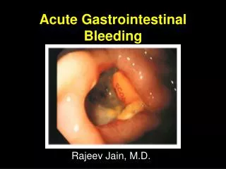
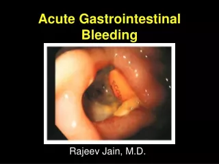
Practical Approach to Acute Gastrointestinal Bleeding Christopher S. Huang MD Assistant Professor of Medicine Boston University School of Medicine Section of Gastroenterology Boston Medical Center
Learning Objectives • UGIB • Nonvariceal (PUD) and variceal • Resuscitation, risk assessment, pre-endoscopy management • Role of endoscopy • Post-endoscopy management • LGIB • Risk assessment • Role and timing of colonoscopy • Non-endoscopic diagnostic and treatment options
Definitions • Upper GI bleed – arising from the esophagus, stomach, or proximal duodenum • Mid-intestinal bleed – arising from distal duodenum to ileocecal valve • Lower intestinal bleed – arising from colon/rectum
Stool color and origin/pace of bleeding • Guaiac positive stool • Occult blood in stool • Does not provide any localizing information • Indicates slow pace, usually low volume bleeding • Melena • Very dark, tarry, pungent stool • Usually suggestive of UGI origin (but can be small intestinal, proximal colon origin if slow pace) • Hematochezia • Spectrum: bright red blood, dark red, maroon • Usually suggestive of colonic origin (but can be UGI origin if brisk pace/large volume)
Case Vignette – CC: • 68 yo male presents with a chief complaint of a large amount of “bleeding from the rectum”
Case Vignette - HPI • Describes bleeding as large volume, very dark maroon colored stool • Has occurred 4 times over past 3 hours • He felt light headed and nearly passed out upon trying to get up to go the bathroom
Case Vignette - HPI • Denies abdominal pain, nausea, vomiting, antecedent retching • No history of heartburn, dysphagia, weight loss • No history of diarrhea or constipation/hard stools • No prior history of GIB • Screening colonoscopy 10 years ago – no polyps, (+) diverticulosis
Case Vignette – PMHx, Meds • Hepatitis C • CAD – h/o MI • PVD • AAA – s/p elective repair 3 years ago • HTN • Hypercholesterolemia • Lumbago • Medications: • Aspirin • Clopidogrel • Atorvastatin • Atenolol • Lisinopril
Case Vignette – Physical Exam • Physical examination: • BP 105/70, Pulse 100, (+) orthostatic changes • Alert and mentating, but anxious appearing • Anicteric • Mid line scar, benign abdomen, nontender liver edge palpable in epigastrium, no splenomegaly • Rectal examination – no masses, dark maroon blood
Case Vignette - Labs • Labs • Hct 21% (Baseline 33%) • Plt 110K • BUN 34, Cr 1.0 • Alb 3.5 • INR 1.6 • ALT 51, AST 76
Initial Considerations • Differential diagnosis? • What is most likely source? • What diagnosis can you least afford to miss? • How sick is this patient? (risk stratification) • Determines disposition • Guides resuscitation • Guides decision re: need for/timing of endoscopy
Differential Diagnosis – Upper GIB • Peptic ulcer disease • Gastroesophageal varices • Erosive esophagitis/gastritis/duodenitis • Mallory Weiss tear • Vascular ectasia • Neoplasm • Dieulafoy’s lesion • Aortoenteric fistula • Hemobilia, hemosuccus pancreaticus Most common Rare, but cannot afford to miss
Differential Diagnosis – Lower GIB Most common diagnosis • Diverticulosis • Angioectasias • Hemorrhoids • Colitis (IBD, Infectious, Ischemic) • Neoplasm • Post-polypectomy bleed (up to 2 weeks after procedure) • Dieulafoy’s lesion
History and Physical History Physical Examination Vital signs, orthostatics Abdominal tenderness Skin, oral examination Stigmata of liver disease Rectal examination Objective description of stool/blood Assess for mass, hemorrhoids No need for guaiac test • Localizing symptoms • History of prior GIB • NSAID/aspirin use • Liver disease/cirrhosis • Vascular disease • Aortic valvular disease, chronic renal failure • AAA repair • Radiation exposure • Family history of GIB
History and Physical History Physical Examination Vital signs, orthostatics Abdominal tenderness Skin, oral examination Stigmata of liver disease Rectal examination Objective description of stool/blood Assess for mass, hemorrhoids No need for guaiac test • Localizing symptoms • History of prior GIB • NSAID/aspirin use • Liver disease/cirrhosis • Vascular disease • Aortic valvular disease, chronic renal failure • AAA repair • Radiation exposure • Family history of GIB
Take Home Point # 1 • Always get objective description of stool • Avoid noninformative terms such as “grossly guaiac positive”
Take Home Point #2 • If you need a card to tell you whether there’s blood in the stool, it’s NOT an acute GIB
Narrowing the DDx: Upper or Lower Source? • Predictors of UGI source: • Age <50 • Melenic stool • BUN/Creatinine ratio • If ratio ≥ 30, think upper GIB J ClinGastroenterol 1990;12:500 Am J Gastroenterol 1997;92:1796 Am J Emerg Med 2006;24:280
Utility of NG Tube • Most useful situation: patients with severe hematochezia, and unsure if UGIB vs. LGIB • Positive aspirate (blood/coffee grounds) indicates UGIB • Can provide prognostic info: • Red blood per NGT – predictive of high risk endoscopic lesion • Coffee grounds – less severe/inactive bleeding • Negative aspirate – not as helpful; 15-20% of patients with UGIB have negative NG aspirate Ann Emerg Med 2004;43:525 Arch Intern Med 1990;150:1381 GastrointestEndosc 2004;59:172
Take Home Point #3 Upper GI bleed must still be considered in patients with severe hematochezia, even if NG aspirate negative
Initial Assessment • Always remember to assess A,B,C’s • Assess degree of hypovolemic shock
Resuscitation • IV access: large bore peripheral IVs best (alt: cordis catheter) • Use crystalloids first • Anticipate need for blood transfusion • Threshold should be based on underlying condition, hemodynamic status, markers of tissue hypoxia • Should be administered if Hgb ≤ 7 g/dL • 1 U PRBC should raise Hgb by 1 (HCT by 3%) • Remember that initial Hct can be misleading (Hct remains the same with loss of whole blood, until re-equilibration occurs) • Correct coagulopathy
Resuscitation • IV access: large bore peripheral IVs best (alt: cordis catheter) • Use crystalloids first • Anticipate need for blood transfusion • Threshold should be based on underlying condition, hemodynamic status, markers of tissue hypoxia • Should be administered if Hgb ≤ 7 g/dL • 1 U PRBC should raise Hgb by 1 (HCT by 3%) • Remember that initial Hct can be misleading (Hct remains the same with loss of whole blood, until re-equilibration occurs) • Correct coagulopathy 40% 40% bleed Time IVFs 20%
Transfusion Strategy • Randomized trial: • 921 subjects with severe acute UGIB • Restrictive (tx when Hgb<7; target 7-9) vs. Liberal (tx when Hgb<9; target 9-11) • Primary outcome: all cause mortality rate within 45 days NEJM 2013;368;11-21
Restrictive Strategy Superior Benefit seen primarily in Child A/B cirrhotics NEJM 2013;368;11-21
Resuscitation • IV access: large bore peripheral IVs best (alt: cordis catheter) • Use crystalloids first • Anticipate need for blood transfusion • Threshold should be based on underlying condition, hemodynamic status, markers of tissue hypoxia • Should be administered if Hgb ≤ 7 g/dL • 1 U PRBC should raise Hgb by 1 (HCT by 3%) • Remember that initial Hct can be misleading (Hct remains the same with loss of whole blood, until re-equilibration occurs) • Correct coagulopathy Weigh risks and benefits of reversing anticoagulation Assess degree of coagulopathy Vitamin K – slow acting, long-lived FFP – fast acting, short lived - Give 1 U FFP for every 4 U PRBCs
Resuscitation • Early intensive resuscitation reduces mortality • Consecutive series of patients with hemodynamically significant UGIB • First 36 subjects = Observation Group (no intervention) • Second 36 subjects = Intensive Resuscitation Group (intense guidance provided) – goal was to decrease time to correction of hemodynamics, Hct and coagulopathy Am J Gastroenterol 2004;99:619
Early Intensive Resuscitation Reduces UGIB Mortality Intervention: Faster correction of hemodynamics, Hct and coags. Time to endoscopy similar (groups are essentially the same) Am J Gastroenterol 2004;99:619
Early Intensive Resuscitation Reduces UGIB Mortality • Observation group • 5 MI • 4 deaths • Intense group • 2 MI • 1 death (sepsis) Am J Gastroenterol 2004;99:619
Causes of Mortality in Patients with Peptic Ulcer Bleeding • Patients rarely bleed to death • Prospective cohort study >10,000 cases of peptic ulcer bleed • Mortality rate 6.2% • 80% of deaths not related to bleeding Am J Gastroenterol 2010;105:84
Causes of Mortality in Patients with Peptic Ulcer Bleeding • Most common causes of non-bleeding mortality: • Terminal malignancy (34%) • Multiorgan failure (24%) • Pulmonary disease (24%) • Cardiac disease (14%) Am J Gastroenterol 2010;105:84
Take Home Point #4 Early resuscitation and supportive measures are critical to reduce mortality from UGIB
Risk Stratification • Identify patients at high risk for adverse outcomes • Helps determine disposition (ICU vs. floor vs. outpatient) • May help guide appropriate timing of endoscopy
Rockall Scoring System • Validated predictor of mortality in patients with UGIB • 2 components: clinical + endoscopic Gut 1996;38:316
AIMS65 • Simple risk score that predicts in-hospital mortality, LOS, cost in patients with acute UGIB Albumin <3.0 INR > 1.5 Mental status altered Systolic BP <90 65+ years old GastrointestEndosc 2011;74:1215
AIMS65 GastrointestEndosc 2011;74:1215
Blatchford Score • Predicts need for endoscopic therapy • Based on readily available clinical and lab data • Can use UpToDate calculator Lancet 2000;356:1318
Blatchford Score GastrointestEndosc 2010;71:1134
Blatchford Score • Most useful for safely discriminating low risk UGIB patients who will likely NOT require endoscopic hemostasis • “Fast track Blatchford” – patient at low risk if: BUN < 18 mg/dL Hgb > 13 (men), 12 (women) SBP >100 HR < 100
Pre-endoscopic Pharmacotherapy • For Non-Variceal UGIB • IV PPI: 80 mg bolus, 8 mg/hr drip • Rationale: suppress acid, facilitate clot formation and stabilization • Duration: at least until EGD, then based on findings
Pre-endoscopy PPI • Reduces the proportion of patients with high risk endoscopic stigmata (“downstages” lesion) • Decreases need for endoscopic therapy • Has not been shown to reduce rebleeding, surgery, or mortality rates High risk Low risk Endoscopic treatment required: Omeprazole – 19% (23% of PUD) Placebo – 28% (37% of PUD) N Engl J Med 2007;356:1631
Endoscopy - Nonvariceal UGIB • Early endoscopy (within 24 hours) is recommended for most patients with acute UGIB • Achieves prompt diagnosis, provides risk stratification and hemostasis therapy in high-risk patients J ClinGastroenterol 1996;22:267 GastrointestEndosc 1999;49:145 Ann Intern Med 2010;152:101
When is Endoscopic Therapy Required? • ~80% bleeds spontaneously resolve • Endoscopic stigmata of recent hemorrhage major
Major Stigmata – Active Spurting Kelsey, PB (Dec 04 2003). Duodenum - Ulcer, Arterial Spurting, Treated with Injection and Clip. The DAVE Project. Retrieved Aug, 1, 2010, from http://daveproject.org/viewfilms.cfm?film_id=39
Adherent Clot • Role of endoscopic therapy of ulcers with adherent clot is controversial • Clot removal usually attempted • Underlying lesion can then be assessed, treated if necessary
Minor Stigmata Flat pigmented spot Clean base Low rebleeding risk – no endoscopic therapy needed
Endoscopic Hemostasis Therapy • Epinephrine injection • Thermal electrocoagulation • Mechanical (hemoclips) • Combination therapy superior to monotherapy Kelsey, PB (Nov 08 2005). Stomach - Gastric Ulcer, Visible Vessel. The DAVE Project. Retrieved Aug, 1, 2010, from http://daveproject.org/viewfilms.cfm?film_id=306 Baron, TH (May 01 2007). Duodenum - Bleeding Ulcer Treated with Thermal Therapy, Perforation Closed with Hemoclips. The DAVE Project. Retrieved Aug, 1, 2010, from http://daveproject.org/viewfilms.cfm?film_id=620
Nonvariceal UGIB –Post-endoscopy management • Patients with low risk ulcers can be fed promptly, put on oral PPI therapy. • Patients with ulcers requiring endoscopic therapy should receive PPI gtt x 72 hours • Significantly reduces 30 day rebleeding rate vs placebo (6.7% vs. 22.5%) • Note: there may not be major advantage with high dose over non-high dose PPI therapy N Engl J Med 2000;343:310 Arch Intern Med 2010;170:751