Download
1 / 17
170 likes | 427 Views
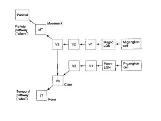
Higher Visual Areas Anatomy of higher visual areas Two processing pathways - “ Where ” pathway for motion and depth - “ What ” pathway for form and color 3. The binding problem. Two anatomical pathways Ventral Pathway:

E N D
Higher Visual Areas • Anatomy of higher visual areas • Two processing pathways • - “Where” pathway for motion and depth • - “What” pathway for form and color • 3. The binding problem
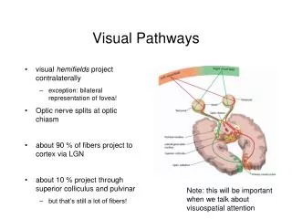
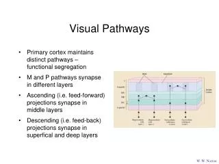
Two anatomical pathways • Ventral Pathway: • Retinal P cells → Parvo LGN → V1 (4Cb) → V2 → V4 → IT (Inferior Temporal Cortex) • 2. Dorsal Pathway: • Retinal M cells → Magno LGN → V1 (4Ca) → V2 → MT (Medial Temporal Cortex) → Posterior Parietal cortex
Ventral Pathway – Two parallel channels for form and color • Parvocellular – interblob system (form): • (V1) L4 (4Cb) → (V1) L2/3 interblob → (V2) pale interstripe → V4 → IT • Parvocellular – blob system (color)x: • (V1) L4 (4Cb) → (V1) L2/3 blob → (V2) thin stripe → V4 → IT
4B (Magno) - Thick stripe Blobs – thin stripe Interblobs - interstripe
Illusory contours can trip firing of V2 cells, while only real contours fire V1 cells.
Temporal Cortex •Inferotemporal Cortex –---Cells respond to a single complex stimulus such as an apple –--- Lesions here leads to inability to identify an object (visual agnosia), picking it up is no problem Superior Temporal Cortex –---Lesions here lead to inability to recognize faces (prospagnosia)
V3 --- Receives input from thick stripe and interstripe areas of V2 --- No thin stripe (Blob) input, generally color insensitive --- Edges of a particular orientation ---Some motion perception --- Depth perception V4 ---Inputs mainly from foveal regions of V1 and V2 (blobs/thin stripes) --- Perceived color of surfaces (not actual wavelengths entering eye) --- Lesions here lead to loss of color vision (Cerebral achromatopsia).,
V5 (MT: Medial temporal cortex) --- Input from thick stripes of V2 (i.e. Magnocellular) ---Specialized for detection of speed and overall motion of entire objects. --- Lesions lead to inability to perceive objects in motion, perception is frozen (Cerebral akinetopsia) l
Agnosias (Sigmund Freud) --Specific defects in vision due to cortical lesion (stroke or tumor). Movement agnosia: Selective loss of movement perception without loss of other perceptual functions, due to bilateral damage in MT or MST Achromatopsia (color agnosia)- loss of color vision due to lesion of temporal cortex (V4) Prosopagnosia – loss of form recognition, due to lesion of inferior temporal cortex
•Complex Cell Responses in Inferior Temporal Cortex 1. Primary cells – respond to simple stimuli 2. Elaborate cells – shapes with color or texture, complex stimuli • 3. Size neurons – invariant neurons: respond to object regardless of size (near or far) - variant neurons: respond to object of a specific size 4. Location neurons – respond to object only in a specific location in the visual field
. Dorsal Pathway - motion and depth Processing Direction-selective Cells – Cells responding to moving bar in one direction, but not in the opposite direction. V1: Many cells with simple and complex RF are direction-selective MT: 1. Direction-selective cells for moving bar or moving dots 2. Columnar organization of direction selectivity 3. RF larger than those of V1 cells 4. Some MT cells (20%) are “pattern direction-selective” 5. Lesion of MT cells impairs motion perception
Aperture Problem Due to small aperture of the receptive field, motion in three directions is perceived as in one direction. Solution: Several lower-order cells project to higher order cells to integrate the local movements.
The binding problem: • ----How the varied aspects of sensory information processed in different cortical areas are integrated to yield the coherent percepts and representations that we experience as the external world. • --- Existence of “Grandmother cell?” • Hypothesis: • Temporal synchrony of neuronal firing may underlie binding. • 2. Cell assembly (Donald Hebb) - The first step of perception is represented by the synchronous firing a specific group of cells. Each cell participate in many different cell assemblies.
Stereopsis -- Perception of solidity or depth for near objects (<100ft) . binocular disparity The difference between the images of an object on the two retinas due to the slightly different location of the two eyes relative to the viewed object. Cues for depth are provided by points just proximal or distal to the fixation point.