Download
1 / 1
10 likes | 241 Views
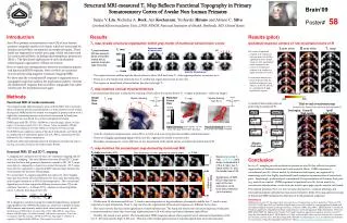
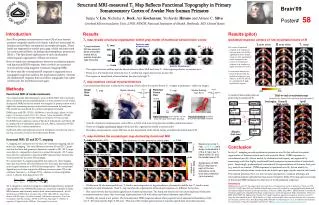
T 1 map reveals structural organization within gray matter of marmoset sensorimotor cortex. T 1 (ms). Posterior ends of lateral sulci. T 1 map (resolution: 0.35 mm isotropic), shown every other coronal slice in posterior to anterior order (#1 to #12). #6. 2050. #1. 1750. #7. #12.

E N D
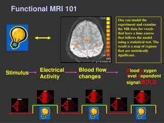
T1 map reveals structural organization within gray matter of marmoset sensorimotor cortex T1 (ms) Posterior ends of lateral sulci T1 map (resolution: 0.35 mm isotropic), shown every other coronal slice in posterior to anterior order (#1 to #12). #6 2050 #1 1750 #7 #12 1450 5 mm Anterior Commissure • The region between midline and the lateral sulcus in slices #5-8 has lower T1. than regions posterior or anterior to it. • There are a few bands with extremely low T1 within this region (red arrows in slice #6). • The region on dorsal bank of lateral sulcus has relatively high T1. T1 map matches cortical myeloarchitecture Coherence Cortical myeloarchitecture is obtained by staining of brain slices for myelin (lower T1 = higher myelination = darker in image). 0.60 0.20 Flattened Cortex View Single Slice View Myelin staining (from Ref.7) Myelin staining T1 map T1 map medial dorsal SI anterior posterior 1 medial lateral 2 1 lateral ventral 2 T1 (ms) Ipsi SII Contra SII 3 3 1875 SII Ipsi SI Low-mye Contra SI Low-mye 1750 2mm X Physical flatteningof cortical gray matter peeled post-mortem Digital flatteningof the slab 0.7-1.3 mm away from gray-white matter boundary. 1625 Ipsi SI High-mye Contra SI High-mye X • Area 3b of primary somatosensory cortex (SI) is a 2 mm wide zone lying between midline and lateral sulcus. • There are 3 highly myelinated bands within area 3b, organized in medial to lateral order. • Secondary somatosensory cortex (SII) lies on the dorsal bank of the lateral sulcus, and abuts the lateral side of SI. Contra Thalamus Stimulus (Left Arm) T1 map (acquired by EPI) 2 mm Slice thickness: 0.5 mm, posterior to anterior order 2000 Based only on the T1 map, cyan lines were drawn to demarcate 2 of the 3 major low-T1 bands within SI, and the medial side of SII. T1 (ms) 1500 Arm electrical stimulation 0.60 Significance of fMRI BOLD response to each of the two stimulation condi-tions was computed (as coherence). FMRI response coherence Leg electrical stimulation 0.20 Structural MRI-measured T1 Map Reflects Functional Topography in Primary Somatosensory Cortex of Awake Non-human Primates Brain’09 Poster#58 Junjie V. Liu, Nicholas A. Bock, Ara Kocharyan, Yoshiyuki Hirano and Afonso C. Silva Cerebral Microcirculation Unit, LFMI, NINDS, National Institutes of Health, Bethesda, MD, United States Introduction Results Results (pilot) Area 3b in primary somatosensory cortex (SI) of non-human primates comprises myelin-rich bands, which are innervated by thalamocortical fibers contralateral to peripheral inputs,. These bands are separated by myelin-poor gaps, which are innervated by corticocortical fibers, including interhemispheric projections (Ref.1). The functional implications of such myelination-related spatial organization of fibers are unclear. Here we study the correspondence between myelination pattern and functional BOLD response, both of which are measured non-invasively using magnetic resonance imaging (MRI). We show that the contralateral SI response is organized into a topographic map that matches the myelination pattern, whereas the ipsilateral SI response does not follow topography but rather centers near the myelination-poor gaps. Ipsilateral response centers on low-myelination band of SI T1 map L arm stim R arm stim The center of ipsilateral response (e.g. response to Left arm stim. in Left SI) is significantly more medial, closer to a low-myelination band, compared to the center of contralateral response (e.g. response to Right arm stim. in Left SI). Co-activation analysis (see below) indicates that the ipsilateral response is not likely to be driven directly by contralateral SI high-myelination regions. Methods Functional MRI of awake marmosets A model of functional pathway projecting to ipsilateral SI Trial-to-trial co-activation map (measures the degree of two regions activating together) Two trained awake adult marmosets were used for fMRI. After received a daily acclimation process and habituated to a sham scanner bore (3 weeks), during each fMRI session the animal was strapped in prone position with a light body-restraining harness, and its head was secured by head posts. The animal was monitored by a camera and appeared relaxed. FMRI scans used EPI, TE/TR = 20/2000 ms, Ernst flip angle, Matrix: 96 (left-right) × 72 (dorsal-ventral). FOV: 32 × 24 mm, 1-shot, bandwidth: 227kHz. Twelve slices (thickness: 0.5 mm). Effective resolution: 0.5 mm isotropic. Each fMRI scan comprises a series of blocks of stimulation; each block (40 s) comprises 8 s of stimulation pulses (1.5 mA, 400 us, repeated at 40 Hz) followed by 32 s without stimulation. Each block delivered unilateral electrical stimulation of either the arm or the leg, via a pair of contact electrodes (diam: 8 mm). Seed region: Contra SI High-myelin Ipsi SI Low-myelin T1 map matches the somatotopic map derived by functional MRI Structural MRI: 2D and 3D T1 mapping T1 mapping was conducted in two ways: 3D volumetric mapping and 2D multi-slice mapping. The only difference between 3D and 2D T1 maps was that the latter had geometric distortions caused by EPI. 3D T1 maps were directly comparable to brain slices stained for myelin. 2D T1 maps were directly comparable to functional MRI response maps because they were measured by the same EPI paradigm. We used a linear T1-mapping algorithm that takes very short imaging time (just 3 inversion times needed) and very short post-processing time, and is largely insensitive to RF/coil inhomogeneities. The conventional algorithm is to fit data acquired at different inversion times (TI) to the nonlinear function S = A+Bexp(-TI/T1), whereas our linear algorithm solves T1 directly from data at the 3 TIs: 3D T1 mapping is conducted using the standard magnetization-prepared rapid gradient-echo (MPRAGE) sequences, which are available on many MRI scanners and routinely used in both clinics and research. Specific details:3 runs of MPRAGE at inversion time of 150, 1400, 4700 ms respectively. Matrix: 108 (left-right) × 90 (dorsal-ventral) × 48 (anterior-posterior). Voxel size (resolution): 0.35 mm isotropic. TE/TR: 2.65/9.3 ms. Flip angle: 9˚. Number of segments: 3. Segment delay: 5000 ms. Nine repeats for each run. Conclusion In-vivo T1 mapping reveals myelination pattern in area 3b that reflects the spatial organization of thalamocortical and corticocortical fibers. FMRI responses in contralateral area 3b, driven mainly by thalamocortical inputs, are organized by somatotopy such that highly myelinated bands comprise representations of major body parts. Surprisingly, ipsilateral and contralateral SI representations of the same body part (e.g. arm) do not match. FMRI responses in ipsilateral area 3b, driven mainly by corticocortical projections, center near the myelin-poor gaps, not the myelin-rich bands. The method presented here is a non-invasive alternative to classical histology and electrophysiological methods that map neural receptive fields. The large spatial coverage of functional MRI facilitates the detection of weak ipsilateral responses. References [1]Krubitzer LA, Kaas JH. The organization and connections of somatosensory cortex in marmosets. J Neurosci 1990; 10(3):952-74. [2] Lipton ML et al. Ipsilateral hand input to area 3b revealed by converging hemodynamic and electrophysiological analyses in macaque monkeys. J Neurosci 2006; 26(1):180-5.[3] Hlushchuk Y and Hari R. Transient suppression of ipsilateral primary somatosensory cortex during tactile finger stimulation. J Neurosci 2006; 26(21):5819-24.[4]Steen RG, Reddick WE, Ogg RJ. Significant regional heterogeneity in human cortical T1. Magn Res Med 2000;18(4):361-8.[5]Eickhoff S et al. High-resolution MRI reflects myeloarchitecture and cytoarchitec-ture of human cerebral cortex. Hum Brain Mapp 2005; 24(3):206-15. [6]Haselgrove J et al. A method for fast multislice T1 measurement. J Magn Res Imag 2000;11(4):360-367. [7]http://udn.nichd.nih.gov/brainatlas_home.html • Within area 3b, the most medial low-T1 band is most responsive to leg stimulation, whereas the middle low-T1 band is most responsive to arm stimulation. Thus T1 map matches the organization of functional responses to different body parts. • The match reveals the functional significance of myeloarchitecture. The hand and the foot are two most important body parts in somatosensation, hence they have large representations in SI with dense myelinated thalamic input fibers. • Notably, the match is not perfect. The hemodynamic fMRI responses almost always spread out of anatomical boundaries of the low-T1 SI bands and the high-T1 SII area. This may reflect larger spatial spread of vascular signals than neuronal responses.