Download

1 / 31
310 likes | 520 Views
Learn about the characteristics, risks, pathology features, clinical presentations, diagnostic criteria, and prognosis of Multiple Myeloma. Understand the differential diagnoses, including MGUS, SMM, Waldenström macroglobulinemia, and solitary plasmacytoma.

E N D
Multiple Myeloma Sujie Tang, MD, MS Hematologist/Oncologist Victory Hematology and Oncology
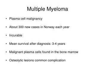
Introduction • Multiple myeloma (MM) is characterized by the neoplastic proliferation of plasma cells producing a monoclonal immunoglobulin. • 1 percent of all cancers and slightly more than 10 percent of hematologic malignancies in the United States
Risk factors The median age at diagnosis is 66 years A small but unknown fraction of cases are familial. The risk of developing MM is approximately 3.7-fold higher for persons with a first-degree relative with MM.
Pathology Features • M- Protein: • IgG – 52 percent; IgA – 21 percent; Kappa or lambda light chain only (Bence Jones) – 16 percent; IgD – 2 percent; Biclonal – 2 percent; IgM – 0.5 percent; Negative – 6.5 percent • Light chain myeloma • Non-secretory myeloma: to be monitored mainly on the basis of imaging tests and bone marrow studies. • Blood Smear: rouleaux formation (>50 percent), leukopenia (20 percent), and thrombocytopenia (5 percent) Bone marrow Biopsy: 10 percent or more clonal plasma cells. Free light chain assay: normal kappa/lambda FLC ratio is 0.26 to 1.65.
Clinical Presentations Signs and symptoms related to the infiltration of plasma cells into the bone or other organs or to kidney damage from excess light chains. ●Anemia – 73 percent • ●Bone pain – 58 percent • ●Elevated creatinine – 48 percent • ●Fatigue/generalized weakness – 32 percent • ●Hypercalcemia – 28 percent • ●Weight loss – 24 percent, one-half of whom had lost ≥9 kg • Less than 5%: • paresthesias (5 percent), hepatomegaly (4 percent), • splenomegaly (1 percent), lymphadenopathy (1 percent), and fever (0.7 percent). • Neurologic disease: cord compression, neuropathy, and CNS involvement
Images • Skeletal surveys: punched-out lytic lesions, diffuse osteopenia, or fractures in nearly 80 percent of patients with MM at the time of diagnosis • CT, MRI, and PET: in patients who have bone pain but no abnormalities on routine xray; and before making the diagnosis of solitary plasmacytoma or smoldering myeloma
Diagnostic Criteria • ●Clonal bone marrow plasma cells ≥10 percent or biopsy-proven bony or soft tissue plasmacytoma PLUS one of the following: • ●Presence of related organ or tissue impairment: CRAB - increased plasma calcium level, renal insufficiency, anemia, and bone lesions. • definitions: • •Anemia – Hemoglobin <10 g/dL (<100 g/L) or >2 g/dL (>20 g/L) below normal • •Hypercalcemia – Serum calcium >11 mg/dL • •Renal insufficiency – Estimated or measured creatinine clearance <40 mL/min (calculator 1 and calculator 2) or serum creatinine >2 mg/dL (177 µmol/liter). • •Bone lesions – One or more osteolytic lesions ≥5 mm in size • ●Presence of a biomarker associated with near inevitable progression to end-organ damage – ≥60 percent clonal plasma cells in the bone marrow; involved/uninvolved free light chain (FLC) ratio of 100 or more; or MRI with more than one focal lesion (involving bone or bone marrow).
DIFFERENTIAL DIAGNOSIS • Monoclonal gammopathy of undetermined significance — Monoclonal gammopathy of undetermined significance (MGUS) is diagnosed in persons who meet the following three criteria: • ●Serum monoclonal protein (whether IgA, IgG, or IgM) <3 g/dL • ●Clonal bone marrow plasma cells <10 percent • ●Absence of lytic lesions, anemia, hypercalcemia, and renal insufficiency (end-organ damage) that can be attributed to the plasma cell proliferative disorder
DIFFERENTIAL DIAGNOSIS • Smoldering multiple myeloma — Smoldering multiple myeloma (SMM) is defined as: • ●M-protein ≥3 g/dL and/or 10 to 60 percent bone marrow plasma cells, plus • ●No end-organ damage or other myeloma-defining events, and no amyloidosis
DIFFERENTIAL DIAGNOSIS • Waldenström macroglobulinemia — lymphoplasmacytic lymphoma (LPL) in the bone marrow • IgM monoclonal gammopathy in the blood • Solitary plasmacytoma — tumors composed of plasma cells of variable maturity, occur solely in the bone, they are designated solitary plasmacytoma of bone. If they arise outside bone in soft tissues, they are called solitary extramedullary plasmacytoma.
Prognosis • Prognosis in myeloma depends on several variables: • 1. host factors (age, performance status, co-morbidities) • 2. disease biology (cytogenetic abnormalities) • 3. stage • 4. response to therapy
Patient factors • Adverse prognostic risk factors for survival (relative risks in parentheses): • ●Performance status 3 or 4 (1.9) • ●Serum albumin <3 g/dL (1.7) • ●Age ≥70 years (1.5) • ●Serum creatinine ≥2 mg/dL (1.5) • ●Platelet count <150,000/microL (1.5) • ●Beta-2-microglobulin >4 mg/L (1.5) • ●Serum calcium ≥11 mg/dL (1.3) • ●Hemoglobin <10 g/dL (1.3) • ●Bone marrow plasma cell percentage ≥50 percent (1.2)
Stage • International staging system (ISS) — • ●Stage I — B2M <3.5 mg/L and serum albumin ≥3.5 g/dL • ●Stage II — neither stage I nor stage III • ●Stage III — B2M ≥5.5 mg/L
Risk Stratification • Based on FISH studies on the bone marrow. If FISH is unavailable, conventional cytogenetics can be used as an alternative but is much less sensitive. • High risk myeloma: median survival of approximately two to three years despite standard treatment • •t(14;16), t(14;20), or del17p13 by FISH • •Elevated lactate dehydrogenase (LDH) levels ≥2 times upper limit of normal • •Features of primary plasma cell leukemia (defined by either ≥2000 plasma cells/microL of peripheral blood or ≥20 percent on a manual differential count) • •High risk signature on gene expression profiling
Risk Stratification • ●Intermediate risk myeloma– Patients with t(4;14) or gain (1q) by FISH or deletion 13/hypodiploidy by conventional cytogenetics were previously considered to have high risk disease, but with appropriate therapy (early use of bortezomib-containing regimens and hematopoietic cell transplantation), patients with these findings have outcomes approaching that of standard risk myeloma. • ●Standard risk myeloma – All patients with MM who lack any of the high or intermediate risk genetic abnormalities described above are considered to have standard risk MM. This includes patients with trisomies, t(11;14), and t(6;14) • With modern therapy, patients with standard risk myeloma have an estimated median survival of 8 to 10 years
Stage • Revised ISS (R-ISS) • ●R-ISS I (n = 871) – ISS stage I (B2M <3.5 mg/L and serum albumin ≥3.5 g/dL) and normal LDH and no del(17p), t(4;14), or t(14;16) by FISH. Estimated OS and PFS at five years were 82 and 55 percent, respectively. Median OS was not reached. Median PFS was 66 months. • ●R-ISS II (n = 1894) – Neither stage I nor stage III. Estimated OS and PFS at five years were 62 and 36 percent, respectively. Median OS and PFS were 83 and 42 months. • ●R-ISS III (n = 295) – ISS stage III (B2M ≥5.5 mg/L) plus LDH above normal limits and/or detection of one of the following by FISH: del(17p), t(4;14), or t(14;16). Estimated OS and PFS at five years were 40 and 24 percent, respectively. Median OS and PFS were 43 and 29 months.
DETERMINING TRANSPLANT ELIGIBILITY Autologous hematopoietic cell transplantation (HCT) compared with chemotherapy alone, intensified chemotherapy followed by HCT appears to prolong both event-free and overall survival in previously untreated patients with standard risk. initial chemotherapy given to patients who are candidates for HCT should avoid agents that may impair stem cell collection Myeloma patients with one or more of the following factors are not considered eligible for autologous HCT in myeloma: • ●Age >77 years • ●Direct bilirubin >2.0 mg/dL (34.2 µmol/liter) • ●Eastern Cooperative Oncology Group (ECOG) performance status 3 or 4 unless due to bone pain • ●New York Heart Association functional status Class III or IV
Regimens and Mechanism • 1. Proteasone Inhibitor: • Bortezomib - proteasome inhibitor . Proteasomes are cellular complexes that break down proteins. The proteins that normally kill cancer cells are broken down too quickly. Bortezomib interrupts this process and lets those proteins kill the cancer cells. • Carfilzomib-Selective proteasome inhibitor. inhibits the chymotrypsin-like activity of the 20S proteasome • Ixazomib - reversibly inhibits the protein proteasome subunit beta type-5 (PSMB5), which is part of the 20S proteasome complex • 2. immunomodulary agent: Thalidamide - anti-angiogenic and oxidative stress-inducing effects. Revlimid (Lenalidomide) - multiple mechanisms of action, three main activities: direct anti-tumor effect, inhibition of angiogenesis, and immunomodulatory role. Pomalidamide- inhibits angiogenesis and myeloma cell growth
Regimens and Mechanism • 3. Daratumumab - It binds to CD38,which multiple myeloma cells overexpress. • 4. Elotuzumab - SLAMF7-directed (also known as CD 319) immunostimulatory antibody • 5. Histone deacetylase inhibitor: • Panobinostat- non-selective histone deacetylase inhibitor • Vornostat : Vorinostat's inhibition of histone deacetylases results in the accumulation of acetylated histones and acetylated proteins, including transcription factors crucial for the expression of genes needed to induce cell differentiation
Choice of Regimens after Relapse • 1. relapse after regimens could replace with newer analogue. • 2. based upon the side effect profile. • 3. An alternative to substituting an existing regimen with new drugs is to add new drugs to the existing regimen. • 4. multi-drug combinations • 5. frail, elderly patients for simpler, well-tolerated regimens such as elotuzumab, lenalidomide, dexamethasone; or ixazomib, lenalidomide, dexamethasone. • 6 Thus the various regimens available should be considered sequentially with each relapse. All categories have been tried at least once in the disease course.
Resources • www.nccn.org • http://www.asco.org/ • http://www.cancer.org/