Download
1 / 27
340 likes | 640 Views
Biological Molecules Nucleic acids and Proteins. Cell Biology Lecture 4. Nucleic acids. Informational macromolecule Deoxyribonucleic acid (DNA) is the genetic material Ribonucleic acid (RNA) Messenger RNA (mRNA) carries information from DNA to the ribosomes

E N D
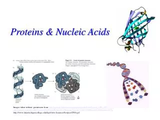
Biological Molecules Nucleic acids and Proteins Cell Biology Lecture 4
Nucleic acids Informational macromolecule Deoxyribonucleic acid (DNA)is the genetic material Ribonucleic acid (RNA) Messenger RNA (mRNA) carries information from DNA to the ribosomes Ribosomal RNA (rRNA) and transfer RNA (tRNA)are involved in protein synthesis RNAs involved in regulation of gene expression and processing and transport of RNAs and proteins
Nucleic acids DNA and RNA are polymers of nucleotides Nucleotidesconsist of Purine and pyrimidine bases Purines: adenine (A) and guanine (G) Pyrimidines:cytosine (C) and thymine(T) RNA has uracil (U)in place of thymine 5 C sugar (5’ phosphorylated) D-Ribose (RNA) D-2’deoxyrobose (DNA) Phosphate: 1-3 phosphate at 5’C of sugar
Nitrogenous bases Structures are meaningful Reactive centers?
Base pairing: Hydrogen bonding Hydrogen bonding btw complementary bases is the basis for double stranded DNA structure
Backbone Sugar phosphodiester forms the backbone Ribose for RNA 2’-deoxyribose for DNA Nucleoside=covalent bonding of C1 of sugar and a base Naming: Guanosine, Adenosine, cytidine and Thymidine, Uridine Nucelotide= Nuceloside+5’phosphate (1-3) Naming: Adenosine monophosphate (AMP) Adenosine diphosphate (ADP) Adenosine triphosphate (ATP) Can you name the others? Adenosine Adenosine monophosphate (AMP) Adenosine diphosphate (ADP) Adenosine triphosphate (ATP)
Phosphodiester bond formation • DNA polymerases catalyze the rxn • uses complementary dNTPs • dehydration reaction between • 3’-OH of new strand and • 5’-phosphate of incoming dNTP • synthesis is 5’3’ • covalent bond is called phosphodiester • there is always a 5’-phosphate and a 3’-OH that gives the DNA its polar sense (5’3’) • complementary strands are anti-parallel
Phosphodiester bond formation • DNA polymerases catalyze the rxn • uses complementary dNTPs • dehydration reaction between • 3’-OH of new strand and • 5’-phosphate of incoming dNTP • synthesis is 5’3’ • covalent bond is called phosphodiester • there is always a 5’-phosphate and a 3’-OH that gives the DNA its polar sense (5’3’) • complementary strands are anti-parallel
DNA is an antiparallel helix • Geometry of bases and their spacial arrangement to form H-bond cause helix structure of dDNA • In B-form right handed dDNA • pairing bases stack in the centre • backbone intertwined • creates minor and major grooves • 0.34 nm (3.4 A) rise per base pair • one full helix turn houses 10 nucleotides Major groove 34 A 20 A
Central dogma • Complementary base pairing allows one strand of DNA to act as a template for synthesis of a complementary DNA or RNA strand • DNA is transcribed to pass genetic information to RNA • The information in RNA is present in a triplet code where every three bases stands for one of the 20 amino acids • Translation: mRNA codes for protein • This flow of information from DNA to protein is called “central dogma” in cell biology • Information flow: DNAmRNAProtein
Central dogma and mutations GAGGUG • The DNA contains the instructions for the sequence of amino acids in each protein • The order of amino acids in a protein determines its shape and function • Errors or faults, ie mutations, in the DNA can change the amino acid sequence and function of the encoded protein • Sickle cell anaemia is due to one nucleotide change affecting hemoglobin reduced O2 carrying capacity
Proteins • Proteinsare the most diverse of all macromolecules • Each cell contains several thousand different proteins • Proteins direct virtually all activities of the cell • Functions of proteins include: • Enzymes • Structural components (e.g. keratin, collagen) • Motility (e.g. actin) • Regulatory (e.g. transcription factors) • Transport (e.g. Na+-K+-ATPase) • Receptors (e.g. insulin receptors) • Transport and storage of small molecules (e.g. O2) • Transmit information between cells (protein hormones), • Defense against infection (antibodies)
Amino acids • Polymers of 20 different amino acids. • Each amino acid consists of the αcarbon bonded to a carboxyl group (COO−), an amino group (NH3+), a hydrogen, and a distinctive side chain (R)
Amino acids • Amino acids are grouped based on characteristics of the side chains: • Nonpolar side chains • Polar side chains • Side chains with charged basic groups • Acidic side chains terminating in carboxyl groups
Nonpolar amino acides • 10 aa have nonpolar R-groups (hydrophobic) • Simplest is glycine (R=H) • 2 contain S and two have cyclic side chains • Nonpolaraa tend to be burried in the hydrophobic core of proteins
Polar amino acides • 5 aa have polar R-groups; either –OH or NH2 (hydrophilic) • Partial charge; H-bond formation with water • Polaraa tend to appear on the surface of proteins
Charged amino acids • 3 aa have positively charged NH2 groups (basic) • Full charge; H-bond and ionic bond • Like Polaraa tend to appear on the surface of proteins • Might take part in catalytic core of enzymes
Charged amino acids • 2 aa have negatively charged –COO- group (acidic) • Full charge; H-bond and ionic bond • tend to appear on the surface of proteins or enzyme catalytic core
Peptide bond formation • Polypeptides: chains of amino acids joined by peptide bonds • Number of aa’s varied • oxytocin – 9 aa, • insulin – 51 aa, titin (connectin)– 34,350 aa’s • Average 400-500 aa • One end of a polypeptide terminates in an αamino group (N terminus) • other end is an αcarboxyl group (C terminus)
Protein structure • Sequence of amino acids in a protein is determined by the order of nucleotide bases in a gene (Primary structure) • One can deduce aa sequence from the sequence of nucleotides in the gene (or mRNA) • 3-D conformation is critical to proteins function • What determines the 3-D structure of proteins?
Protein secondary structure Christian B. Anfinsen (1957) • 3-D structure is a result of interactions between the amino acids • Christian Anfinsen denaturedribonuclease (RNase) by heat treatment; breaks H-bonds • If the treatment was mild, the proteins would return to their normal shape at room temperature • This would mean that the information for folding the protein is in its primary sequence (how could he test?)
Protein secondary structure • Secondary structure: regular arrangement of amino acids within localized regions • There are 2 types of secondary structure: • The polypeptide can coil in a spiral helix shape • The polypeptide can fold to form a βpleated sheet (parallel or antiparallel) • Both are held together by hydrogen bonds between the CO and NH groups of peptide bonds
Protein Tertiary structure • Observation: • Similarly disrupting the disulfide bonds (S-S) using chemical denaturing agents (eg. β-mercaptoethanol) denatures proteins (-SH forms) • Incubation under oxygen refolded the RNase back to its functional conformation (ie enzyme gained capacity to degrade RNA) • indicates a higher level of structure important for function that relies on covalent S-S bridge (tertiary structure)
Protein Tertiary structure • Tertiary structure: folding of secondary structural elements to form a 3-D arrangement • 2° elements connected by loops and less ordered aa’s • interactions btw the side chains of amino acids in different regions of protein stabilizes the 3° structure • Covalent bonds (S-S bridge) • Hydrophobic and hydrophilic interactions • In most proteins this results in domains, the basic units of tertiary structure RNase Insulin
Protein Quaternary structure • Quaternary structure consists of interactions between different polypeptide chains • In multi-subunit enzymes • Hemoglobin, for example, is composed of four polypeptide chains
Protein structure: Summary Campbell & Reece, 2002
Can you meet these objectives? • Distinguish among nucleosides, nucleotides and nucleic acids? • Explain the structure of DNA? • List some functions of proteins in cells? • Describe and distinguish between amino acids? • Discuss the levels of protein structure and organization of proteins?