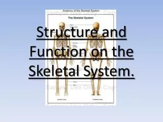
STRUCTURE AND FUNCTION OF THE NEUROLOGIC SYSTEM
430 likes | 548 Views
STRUCTURE AND FUNCTION OF THE NEUROLOGIC SYSTEM. Organization. Central nervous system (CNS) Anatomical structures Brain enclosed -- cranial vault Spinal cord enclosed -- bony spine Peripheral nervous system (PNS) Anatomical organization (Fig.12-26) Nerves Cranial – 12 pairs

STRUCTURE AND FUNCTION OF THE NEUROLOGIC SYSTEM
E N D
Presentation Transcript
Organization • Central nervous system (CNS) • Anatomical structures • Brain enclosed -- cranial vault • Spinal cord enclosed -- bony spine • Peripheral nervous system (PNS) • Anatomical organization (Fig.12-26) • Nerves • Cranial – 12 pairs • Spinal -- 31 pairs • Can be afferent or efferent
Functional organization • Somatic nervous system • Regulates voluntary, motor control • Neurotransmitter = acetyl choline (ACh) • Autonomic nervous system • Regulates internal environment
Most organs dually innervated: • Sympathetic neurons (Fig.12-24,23) • From thoracic, lumbar spinal regions • Important for “fight or flight” (incr’d heart rate/resp’n, decr’d digestion) • Neurotransmitters: ACh and epinephrine/norepi
Parasympathetic neurons (Fig.12-25,23) • From other spinal regions • Important to conserve energy and maintain homeostasis (decr’d heart rate, incr’d digestion) • Neurotransmitter: ACh
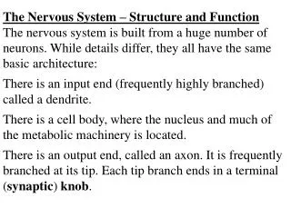
Neural tissue • Neuron = primary cell of nervous system • About 1011 neurons/body • Each neuron adapted for specific function • Functions of neurons • Detect env’l changes • Initiate body response to changes • Fuel source -- mostly glucose
Anatomic components (Fig.12-1) • Cell body = soma • Most in CNS • Those in PNS grouped together as ganglia • Dendrites • Extensions of cell body • Carry information TOWARD cell body
Axon • Usually one per neuron • Long projection; carries impulses AWAY from cell body • Myelin – insulating lipid covering • Forms sheath • Allows fast flow of ions in one direction proper impulse conduction (away from cell body) • Interruptions in myelin coating = nodes of Ranvier • Nec for ions from ISF to enter axon for proper impulse
Supporting cells of neurological system (Fig.12-3,Table12-1) • Schwann cells – in PNS • Form myelin sheath around axons • Neuroglia -- “nerve glue” • Support CNS neurons • About ½ volume of the brain and spinal cord
Several types of neuroglial cells: • Astrocytes -- star shape • Form contact between neurons, circulatory system • “Buffer zone” between neurons (delicate) and molecules circulating in blood • Oligodendroglia • Deposit myelin in CNS (similar job as the Schwann cells in PNS) • Microglia • Phagocytic cells; digest debris in CNS • Ependymal cells • Help produce cerebrospinal fluid (csf)
Nerve injury and regeneration (Fig.12-4) • Mature neurons don’t divide, proliferate • Injury can permanent loss of function • Regeneration of some PNS neurons is possible • Axon of neuron (so only myelinated fibers) repaired • Regeneration more optimistic if cell crushed • If cut, scar tissue can form impede ion flux through cell membrane, so impede proper impulses
Regeneration more optimistic if injury further away from cell body • With regeneration, see: • Swelling distal to injury • Filaments hypertrophy • Myelin sheath and axon begin to degenerate, BUT • Proximal to injury, see projection of new neurofibriles • Neurilemma (membrane that surrounds the myelin sheath) acts as guide • Not in CNS, where myelin somewhat different • Scar tissue forms, and decr’d/no regeneration of neuronal tissue
Nerve impulses • Action potentials generated • Neuron selectively changes electrical potential of its plasma membrane • Influx of Na+ through selective channels (gated Na+ channels) at dendrite or soma • In response to biochemical signal from a neurotransmitter released from an impinging neuron • Changes electrical potential of membrane in that region
Neurons influence neighboring neurons (Fig.12-2) • Release neurotransmitters (biochemicals signal an action potential in a neighboring neuron) • Synapse – region between two nearby neurons • First neuron in a series = “presynaptic” • Second neuron =“postsynaptic” • Presynaptic impinges on postsynaptic • Neurotransmitters synth’d, stored in vesicles near end of presynaptic neuron
When action potential reaches end of presynaptic neuron: • Signals vesicle holding neurotransmitters to merge with neuron’s plasma membrane in presynaptic area • Neurotransmitters released into synapse • Neurotransmitters travel through synapse, where they encounter postsynaptic neuron • On plasma membrane of postsynaptic neuron is a receptor specific for a particular neurotransmitter
Neurotransmitter binds the receptor on the postsynaptic neuron • Signals opening of nearby Na+ channels • Membrane potential changes in the postsynaptic neuron • Generation of action potential • Action potential travels through postsynaptic neuron’s dendrite, cell body and axon to axon ending (now presynaptic) • Signals neurotransmitter release to next neuron or muscle fiber on which it impinges, and changes occur within that cell
Some widely studied neurotransmitters • Norepinephrine, epinephrine, dopamine, ACh, serotonin (and MANY others)(Table12-2) • Excitatory neurotransmitters cause Na+ to flood into neuron depolarization and action potential • Inhibitory neurotransmitters dampen Na+ influx into neuron inhibition of depolarization, so no action potential • Different neurotransmitters have different functions (some excitatory, some inhibitory)
Central Nervous System (CNS) • The brain • Allows reasoning, intelligence, personality, mood • Weighs about 3 lb. in average adult • Receives about 20% of cardiac output • Divisions (Table 12-3;Fig.12-6) • Different regions, each associated with different function (Fig.12-7) • BUT some functions controlled by more than one region • Ex: cerebrum -- centers for sensory/motor, reasoning, memory, intelligence
Characteristics/Structures • Gyri – convolutions of tissue along brain surface • Importance: increase surface area of brain • Sulci – grooves between gyri • Gray matter – cerebral cortex • Cell bodies of neurons (so not myelinated) • White matter – myelinated nerve fibers (= axons) • Lies beneath cerebral cortex
Spinal cord (Fig.12-9,10,11) • Long nerve cable • Continuous with brain • Lies in vertebral canal • Surrounds, protects spinal cord • Divided into 31 anatomical sections • Gray matter (Fig.12-11) • In center of spinal cord • Butterfly shaped • Divided 3 horns • Composed of neuronal cell bodies
White matter • Surrounds gray matter • Myelinated tissue (so axons) • Forms ascending, descending tracts • Motor neurons (Fig.12-12,13) • Directly influence the muscle cells • Cell bodies of motor neurons lie in gray matter of spinal cord • Axons extend out of spinal cord • Regulate motor activity
Protective structures of the CNS • Cranium • 8 fused bones; encloses and protects the brain • Epidural space • Lies between cranium and meninges • Site of blood collection ( epidural hematoma) if trauma disruption of blood vessels of scalp/skull
Meninges – 3 protective membranes (Fig.12-14): • Dura mater – 2 layers of tissue • Arachnoid membrane – named for appearance (spider web) • Pia mater – cells to produce cerebrospinal fluid • Spaces between layers -- also sites where blood may collect if hemorrhage
Cerebrospinal fluid (csf) • Clear, colorless fluid similar to ISF and plasma (Table 12-4) • Helps cushion CNS • Produced within pia mater (about 600 mL/day) • Circulates within cranium in cavities, subarachnoid space • Exerts pressure within brain, spinal cord • Forms pressure gradient between arteries, cavities of CNS • Reabsorbed into venous circulation • Valves in arachnoid membrane move fluid into venous circulation (and opposite)
Vertebral column (Fig.12-15,16) • Vertebrae • 33 • Intervertebral discs • Between vertebrae • Pulpy, absorb shock • Prevent damage to nervous system structures • If rupture back pain
Vertebral circulation • Arises from aortic arch internal carotid arteries and vertebral arteries (Fig.12-18; Table 12-5) • May be conducting ( brain surface), OR • Penetrating ( structures below the cortex) • Healthy brain can regulate its blood supply to maximize oxygen supply • Can increase extraction of oxygen from blood when systemic bp decreases (for awhile) • Can decrease resistance in cerebral vessels when systemic bp decreases (up to a point)
Blood-brain barrier • Supporting neural cells and blood capillaries have rel. tight junctions • Selectively allow partic blood components from blood brain • Important in brain chemotherapy