
AKNOWLEDGEMENTS
E N D
Presentation Transcript
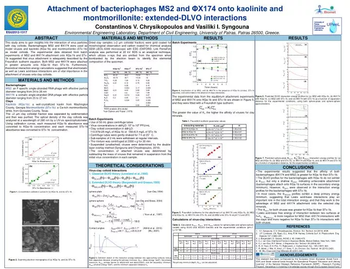
Attachment of bacteriophages MS2 and ΦX174 onto kaolinite and montmorillonite: extended-DLVO interactions Constantinos V. Chrysikopoulos and Vasiliki I. Syngouna Environmental Engineering Laboratory, Department of Civil Engineering, University of Patras, Patras 26500, Greece. EGU2012-1317 ABSTRACT MATERIALS AND METHODS RESULTS RESULTS This study aims to gain insights into the interaction of virus particles with clay colloids. Bacteriophages MS2 and ΦX174 were used as model viruses and kaolinite (KGa-1b) and montmorillonite (STx-1b) as model colloids. The experimental data obtained from batch experiments of MS2 and ΦX174 attachment onto KGa-1b and STx-1b suggested that virus attachment is adequately described by the Freundlich isotherm equation. Both MS2 and ΦX174 were attached in greater amounts onto KGa-1b than STx-1b. Furthermore, theoretical interaction energy calculations suggested that electrostatic as well as Lewis acid-base interactions are of vital importance in the attachment of viruses onto clay colloids. • Dried clay samples (<2 μm colloidal fraction) were gold coated for morphological observation and carbon coated for chemical analysis SEM (JEOL-6300 microscope) with EDS (OXFORD, Link PentaFet) analysis was performed at 20 kV. EDS is an analytical technique, which utilizes x-rays that are emitted from the specimen when bombarded by the electron beam to identify the elemental composition of the specimen. • Batch Experiments • Use of 50 mL glass centrifuge tubes • Virus concentrations in ddH2O: 103 to 109 PFU/mL • Clay colloid concentration in ddH2O: • 113.57925 mg/L of KGa-1b or 1606.5 mg/L of STx-1b • Centrifuge tubes were gently shaked for 7 h at 25°C. • Sub-samples of 2 mL were withdrawn at regular intervals. • The mixture was centrifuged at 2000 x g for 30 min. • Suspended (unattached) viruses were determined by the double-layer overlay method (Syngouna and Chrysikopoulos, 2010). • The concentration of attached viruses was determined by subtracting the mass of viruses that remained in suspension from the initial virus concentration in each sample. Batch Experiments MATERIALS AND METHODS Bacteriophages MS2: an F-specific single-stranded RNA phage with effective particle diameter ranging from 24 to 26 nm ΦX174: a somatic single-stranded DNA phage with effective particle diameter ranging from 25 to 27 nm Clays Kaolinite (KGa-1b): a well-crystallized kaolin from Washington County, Georgia Montmorillonite (STx-1b): a Ca-rich montmorillonite, white, from Gonzales County, Texas The <2 μm clay colloidal fraction was separated by sedimentation and then was purified. The optical density of the clay colloids was analyzed at a wavelength of 280 nm by a UV-vis spectrophotometer. Using calibration curves, each measured KGa-1b absorbance was converted to KGa-1b concentration and each measured STx-1b absorbance was converted to STx-1b concentration. Figure 4.Inactivation of (a) MS2, and (b) ΦX174 in the presence of KGa-1b (circles), STx-1b (squares), and control data in the absence of clays (diamonds). The experimental data from the equilibrium attachment experiments of MS2 and ΦX174 onto KGa-1b and STx-1b are shown in Figure 5, and they were fitted with a Freundlich type isotherm: The greater the value of Kf, the higher the affinity of viruses for clay minerals. Table 1. Freundlich isotherm parameter values Figure 6. Predicted DLVO interaction energy profiles for (a) MS2 with KGa-1b, (b) ΦX174 with KGa-1b, (c) MS2 with STx-1b, and (d) ΦX174 with STx-1b as a function of separation distance for the experimental conditions, using both sphere-plate and sphere-sphere approximations. †EDS analysis (this study) ‡van Olphen and Fripiat (1979) Figure 7. Predicted sphere-plate ΦDLVO, ΦAB, and ΦXDLVO interaction energy profiles for (a) MS2 and KGa-1b, (b) MS2 and STx-1b, (c) ΦX174 and KGa-1b, and (d) ΦX174 and STx-1b as a function of separation distance, for the experimental conditions. THEORETICAL CONSIDERATIONS CONCLUSIONS Virus-clay colloid interactions 1. Classical DLVO theory (Loveland et al.,1996) 2. Extended DLVO theory (Bergendahl and Grasso,1999) sphere-plate (van Oss,1994) sphere-sphere (van Oss and Giese, 2004) , ( Yoon et al., 1997) Contact angles: MS2=331, X174=261.7 (Attinti et al., 2010) KGa-1b=46.1, STx-1=20.52.8 (Wu, 2001) • The experimental results suggested that the affinity of both bacteriophages (ΦX174 and MS2) is greater for KGa-1b than STx-1b. • The DLVO profiles for the bacteriophages with KGa-1b do not exhibit a min1, but only a shallow min2, indicating unfavorable attachment (bacteriophages attach onto KGa-1b surfaces in the secondary energy minimum). However, min1were observed in the interaction energy profiles for the bacteriophages with STx-1b. • In most cases, the XDLVO profiles exhibit a deep primary energy minimum, suggesting that Lewis acid-base interactions play an important role in the total interaction energy, and that they work to the advantage of MS2 and X174 attachment onto the selected clay minerals. • The min1 for both viruses was greater for KGa-1b than STx-1b. • Lewis acid-base free energy of interaction between two surfaces at h=ho, is more negative for MS2 than X174 interactions with the clays and more negative for KGa-1b than STx-1b interactions with both viruses. Figure 1. Concentration calibration curves for (a) KGa-1b, and (b) STx-1b. Figure 5. Freundlich isotherms for the attachment of (a) ΦX174 onto KGa-1b, (b) MS2 onto KGa-1b, (c) ΦX174 onto STx-1b, and (d) MS2 onto STx-1b at pH 7.0 and 25oC. Calculations of virus-clay interactions Table 2. Calculated max1,, min1, and min2 values for sphere-plate and sphere-sphere models using DLVO and XDLVO theories and the experimental conditions (pH=7, Is=10-4 M). †No primary minimum depth, min1 can be calculated. REFERENCES • V.I. Syngouna, C.V. Chrysikopoulos, Environ. Sci. Technol. 44 (2010) 4539. • J.P. Loveland, J.N. Ryan, G.L. Amy, R.W. Harvey, Colloids Surf. A: Physicochem. Eng. Aspects 107 (1996) 205. • J. Bergendahl, D. Grasso, AIChE J. 45 (1999) 475. • C.J. van Oss, Interfacial Forces in Aqueous Media, Marcel Dekker, New York, 1994. • C.J. van Oss, R.F. Giese, J. Dispersion Sci. Technol. 25 (2004) 631. • R.-H. Yoon, D.H. Flin, Y.I. Rabinovich, J. Colloid Interf. Sci. 185 (1997) 363. • R. Attinti, J. Wei, K. Kniel, J.T. Sims, Y. Jin, Environ. Sci. Technol. 44 (2010) 2426. • W. Wu, Clays and Clay Minerals 49 (2001) 446. AKNOWLEDGEMENTS Figure 3. Definition sketch of the interaction energy between two approaching surfaces versus their separation distance, showing the primary minimum, min1 (deep energy "well"), the primary maximum, max1 (energy barrier to attachment and detachment), and the secondary minimum, min2 (shallow energy "well"), and the minimum separation distance ho. This research has been co-financed by the European Union (European Social Fund – ESF) and Greek national funds through the Operational Program "Education and Lifelong Learning" of the National Strategic Reference Framework (NSRF) - Research Funding Program: Heracleitus II. Investing in knowledge society through the European Social Fund. Figure 2.Scanning electron micrographs of (a) KGa-1b, and (b) STx-1b.