Download
1 / 17
170 likes | 281 Views
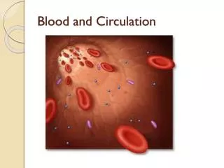
Circulation and Heart Structures. Unit D – Human Systems. Circulatory Systems in your Body. There are two circulatory systems in your body: Pulmonary circulatory system Systemic circulatory system. 1. Pulmonary Circulatory System.

E N D
Circulation and Heart Structures Unit D – Human Systems
Circulatory Systems in your Body • There are two circulatory systems in your body: • Pulmonary circulatory system • Systemic circulatory system
1. Pulmonary Circulatory System • Blood vessels that circulate blood between the heart and the lungs. • Carries deoxygenated blood to the lungs and brings oxygenated blood back to the heart.
2. Systemic Circulatory System • Blood vessels that carry blood between your heart and all other parts of the body. • Pumps oxygenated blood to all body tissues and returns deoxygenated blood back to the heart. • Watch this: http://www.youtube.com/watch?v=0jznS5psypI
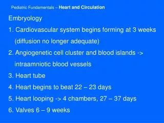
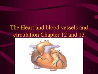
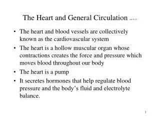
Basic Heart Anatomy • The heart is made up of two halves, the left and the right. • Each side of the heart is comprised of two chambers. • Upper chambers are called atria, lower chambers are called ventricles.
Starting with deoxygenated blood coming from the vena cava • Deoxygenated blood from your head and upper body enters the right atrium of your heart from the superior vena cava. • Deoxygenated blood from the lower regions of your body enters the right atrium of your heart from the inferior vena cava.
Events occurring in the right atrium • Blood collects in the right atrium until the pressure inside forces a set of valves called the right atrioventricular (AV)/tricuspid valves open. • Valves make sure that blood only travels in one direction. • Blood now enters the right ventricle, where it pools until the pressure inside increases, forcing the semilunar valves open.
Blood flow to the lungs • Semilunar valves separate the ventricles from the arteries. • Deoxygenated blood now flows through the left and right pulmonary arteries to the lungs. • In the lungs, carbon dioxide will be released, and oxygen will combine with hemoglobin.
Blood flow back to the heart • Oxygenated blood returns to the heart via the left and right pulmonary veins, where they will empty into the left atrium of the heart. • Blood will pool in the left atrium of the heart, until the pressure builds up, forcing the left AV (bicuspid) valves to open.
Blood flow to the body • Blood passes through the left AV valves into the left ventricle. • Again, blood collects until sufficient pressure builds up. • Blood passes through the left semilunar valves into the aorta, the largest artery in your body. • The aorta branches off into smaller arteries, taking blood to all parts of your body.
Heart Muscle • Made of myogenic muscle. • Myogenic muscle has the ability to contract without stimulation from the nervous system…it can beat by itself. • For a short time, your heart will continue to beat, even if it is removed from the body. • Watch this: http://www.youtube.com/watch?v=iX6HnUyzgQ0&NR=1
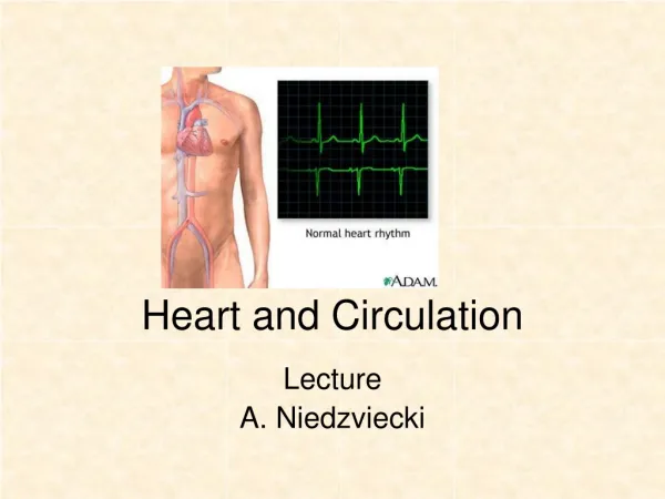
Heart Rate and Contractions • Set by the sinoatrial (SA) node, a bundle of nerves known as the “pacemaker” of the heart. • Heart rate is typically set at about 70 beats per minute. • SA node sends nerve impulses to another bundle of nerves called the atrioventricular (AV) node. • Watch this: http://www.youtube.com/watch?v=Vq0_5RL7cUk&NR=1
This nervous impulse causes the atria of the heart to contract, pushing blood into ventricles. • The signal is then continued to the end of the ventricles causing them to contract, pushing the blood into the arteries.
Diastole • Relaxation of heart muscle, when the atria of the heart are filling with blood. • Increased blood volume and muscle contraction increase blood pressure, forcing the AV valves open. • Blood rushes into the ventricles of the heart, causing the AV valves to shut. • This causes the heavy “LUBB” sound.
Systole • Very quickly, increase blood volume and muscle contractions increase pressure in the ventricles. • This forces semilunar valves open, letting blood rush into arteries. • Semilunar valves close, causing the lighter “DUBB” sound.
Heart Murmur • Occurs when heart valves do not close properly. • Can be diagnosed by hearing a gurgling sound when listening with a stethoscope. • This means that blood can flow backwards, not in the direction it is intended to. • Decreases oxygen delivery to body tissues.
Student Tasks for Lesson • Label Heart Structures diagram given to you by your teacher and colour parts of heart accordingly: red for parts carrying oxygenated blood, blue for parts carring deoxygenated blood. • Complete #1-3, 7 on page 327.