Download
1 / 73
1.06k likes | 2.62k Views
Leptospirosis update. Dr.T.V.Rao MD. Scientific Begin ning. It was first described by Adolf Weil in 1886 when he reported an "acute infectious disease with enlargement of spleen, jaundice and nephritis". Leptospira was first observed in 1907 from a post mortem renal tissue slice . .

E N D
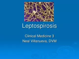
Leptospirosis update Dr.T.V.Rao MD Dr.T.V.Rao MD
Scientific Beginning • It was first described by Adolf Weil in 1886 when he reported an "acute infectious disease with enlargement of spleen, jaundice and nephritis". Leptospira was first observed in 1907 from a post mortem renal tissue slice. Dr.T.V.Rao MD
Leptospirosis - Zoonosis • Leptospirosis is an acute arthropod-zoonotic infection of worldwide significance caused by spirochete Leptospira interregna's which has 23 serogroups and >200 serovars. Various factors influencing the animal activity, suitability of the environment for the survival of the organism and behavioural and occupational habits of human beings can be the determinants of incidence and prevalence of the disease. Dr.T.V.Rao MD
What is leptospirosis? • Leptospirosis, also known as canicola fever, haemorrhagic jaundice, infectious jaundice, mud fever, spirochetal jaundice, swamp fever, swineherd's disease, caver's flu or sewerman's flu, is a bacterial infection resulting from exposure to the Leptospira interrogans bacterium. Dr.T.V.Rao MD
Leptospirosis also called as Weil’s Disease after its inventor . Dr.T.V.Rao MD
Weil’s disease signifies Leptospirosis • There is an acute form of human infection known as Weil's disease, where the patient suffers from jaundice, though this term is often (incorrectly) used to describe any case of infection.. Dr.T.V.Rao MD
Leptospirosis 2011 Dr.T.V.Rao MD
Synonyms Dr.T.V.Rao MD
The Causative Bacterium Dr.T.V.Rao MD
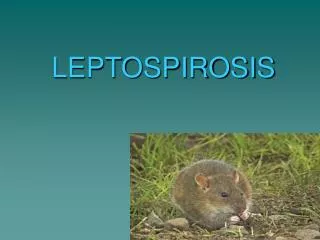
Reservoirs • Wild and domestic animals rodents, livestock (cattle, horses, sheep, goats, swine), canines, and wild mammals are the reservoir for leptospirosis. Many animals have prolonged leptospiruria without suffering from the disease themselves. Dr.T.V.Rao MD
Classification: • Phylum: Spirochaetes • Class: Spirochaetes • Order: Spirochaetales • Species: Leptospira • Family: Leptospiraceae Dr.T.V.Rao MD
What causes Leptospirosis • Leptospirosis is a bacterial disease that affects humans and animals. Leptospira bacteria are found worldwide and there are many different types or serovars capable of causing disease. Disease caused by Leptospira bacteria is most common in temperate or tropical climates and appears to be rare in North America. Dr.T.V.Rao MD
Morphology • The Leptospira appear tightly coiled thin flexible Spirochetes 5 – 15 microns long. • Fine spiral of 0.1 – 0.2 microns • One end appears bent forms a hook. • Actively motile • Seen best with dark field Microscopy. Dr.T.V.Rao MD
Greater Understanding with Electron Microscopy • Electron Microscopy show thin axial filament and a delicate membrane • In dark field it may appear as chain of miniature cocci. Dr.T.V.Rao MD
Resistance and Disinfection • Leptospira species can be inactivated by 1% Sodium hypochlorite • 70%ethanol, • glutaraldehyde, • formaldehyde, • detergents and acid. • This organism is sensitive to moist heat (121 ° C for a minimum of 15 min))and is also killed by pasteurization. Dr.T.V.Rao MD
Leptospirosis – A Major Zoonotic Infection • Weil's disease is comparatively rare, though 'mild' cases of leptospirosis happen everywhere there are carriers, and it is believed that leptospirosis is one of the most common zoonotic infections in the world. Millions of people are infected each year, but information and treatment can be limited, especially in the developed world where cases are considered 'rare' by the medical community. Dr.T.V.Rao MD
Animals spread Leptospirosis Rats, Mice, Wild Rodents, Dogs, Swine, Cattle are principle source of infection The above animals excrete Leptospira both in active infection and Asymptomatic stage The Leptospira survive and remain viable for several weeks in stagnant water. Dr.T.V.Rao MD
Modes of Transmission 1. Direct contact with urine or tissue of infected animal Through skin abrasions, intact mucus membrane 2. Indirect contact Broken skin with infected soil, water or vegetation Ingestion of contaminated food & water 3. Droplet infection Inhalation of droplets of infected urine Dr.T.V.Rao MD
Transmission Human infection is accidental No human to human transmission Dr.T.V.Rao MD
Pathogenic Strains x Non pathogenic Leptospirosis • There are several species of Leptospira only few are pathogenic to Humans, rest to some Animals and Many in Nature as saprophytes • Leptospira Interrogans is Pathogenic there are 200 serovars. • Leptospira biflexa Non Pathogenic there are 60 serovars • Further classifications are made on shared antigens Dr.T.V.Rao MD
Genomic based classification • DNA – DNA hybridization studies proved more specific • The traditional serologic classification has limitations at Molecular level, but useful at Epidemiological studies. Dr.T.V.Rao MD
Comparative Morphology of Spirochetes Dr.T.V.Rao MD
Culturing of Leptospira • Leptospira grows best under aerobic conditions at 280 to 300c best demonstrated in Semisolid agar media • Optimal Media Fletchers Media Stuarts Media Optimal growth after 1 – 2 weeks Dr.T.V.Rao MD
Growth requirements • Leptospira derive energy from oxidation of long chain fatty acids, and cannot use or carbohydrates or amino acids as major energy source. Dr.T.V.Rao MD
Antigenic structure • All isolates of L.inttterogans from different parts of the world are serologically related and exhibit cross reactions in serologic tests. • Overlapping of Antigens do occur in different species. • Outer envelop contains large amount of Lipopolysaccharides ( LPS ) • Antigenic structure varies from one strain to other • This variation forms the basis of serologic classification Dr.T.V.Rao MD
Genome of Leptospira • L. interrogans serogroups Icterhaemorrhagiae consists of a 4.33 mega base large chromosome and a 359 kilo base small chromosome, totalling 4,768 predicted genes. A series of genes have been discovered that could potentially be related to adhesion. This genome differs from the two other pathogenic spirochete (Treponema palladium and Borrelia burgdorferi), though some similar genes are visible (CHGC, 2004). Dr.T.V.Rao MD
PATHOGENESIS • leptospira • skin,mucosa • Initial stage leptospiremia toxic symptoms • (1~3days) three symptoms: • fever,myalgia,fatigue; • three signs: • conjunctival suffussion; • muscle tenderness; • enlargement of lymphonodes; Dr.T.V.Rao MD
Pathogenesis • Leptospira are present in the water bodies • Enter through breaks in the skin ( cuts and abrasions ) and mucous membranes • Enters through Mouth – Nose – Conjunctive • Rarely enters though ingestion. • Incubation period 1 – 2 weeks • When multiples blood stream produces fever. • May establish organ involvement in Kidney and Liver, • May produce hemorrhage and necrosis in the tissues and initiates dysfunction of these organs Dr.T.V.Rao MD
Sequence of Leptospira Infection Dr.T.V.Rao MD
Clinical Presentation Dr.T.V.Rao MD
May present with • Jaundice • Hemorrhage • Nitrogen retention • The Illness is Biphasic with initial temperature when the second phase comes with raise of IgM titers raise • Aseptic meningitis – initial headache, stiffness of neck, pleocytosis of Cerebro spinal fluid Dr.T.V.Rao MD
Pathogenesis of Severe Disease Vasculitis Damage to small blood vessels Leptospira Massive migration of fluid from Intravascular to interstitial compartment Direct cytotoxic injury Immunological injury Renal dysfunction, vascular Injury to internal organs Dr.T.V.Rao MD
Presenting with Jaundice is significant and Important, Serious Manifestation Dr.T.V.Rao MD
May present with Major Complications • Nephritis • Hepatitis. • Manifestations in eye • Muscular lesions • Many infections are mild and subclinical Dr.T.V.Rao MD
Weil’s Syndrome • Weil's syndromeis a severe form of leptospirosis that causes a continuous fever, stupor, and a reduction in the blood's ability to clot, which leads to bleeding within tissues. Blood tests reveal anaemia. By the third to sixth day, signs of kidney damage and liver injury appear. Kidney abnormalities may cause blood in the urine and painful urination. Liver injury tends to be mild and usually heals completely. Dr.T.V.Rao MD
May present as Atypical Pneumonia Dr.T.V.Rao MD
Hepatitis - Leptospirosis • Hepatitis is the frequent complication • Elevation of serum creatine phospholipase enzyme raise differentiates from Viral hepatitis where the enzyme is not raised Dr.T.V.Rao MD
Nephritis - Leptospirosis • Kidney involvement in animals produce chronic disease of the kidney and the infected animal starts shedding large number of Leptospira and main source of environmental contamination of bacteria and results I human infections • Human urine also contain Spirochetes in the second and third week of infection Dr.T.V.Rao MD
Complications • Azotemia • Oliguria • Hemorrhage • Purpura • Hemolysis • Gastrointestinal bleeding • Hypoprothrombinemia and Thrombocytopenia Dr.T.V.Rao MD
Early and Prompt Diagnosis is Highly Essential • The development of simpler, rapid assays for diagnosis has been based largely on the recognition that early initiation of antibiotic therapy is important in acute disease but also on the need for assays which can be used more widely. Dr.T.V.Rao MD
Laboratory Tests • TC / DC / ESR / Hb / Platelet count • Serum Bilirubin / SGOT/ SGPT • Blood Urea, Creatinine & Electrolytes • Chest X-Ray; ECG • Tests for diagnosis of Leptospirosis • Culture for Leptospira: Positive • MAT; Sero conversion or 4 fold rise/ high titer • ELISA / MSAT : positive • MAT: Microscopic agglutination test • (M)SAT: Microscopic slide agglutination Test
Approach to Diagnosis Dr.T.V.Rao MD
Laboratory Diagnosis Specimens 1 Blood to be collected in a heparin tube 2 CSF, Tissues Microscopic examination 3 Urine to be collected with great care to avoid contamination 4 Serum for agglutination tests Dr.T.V.Rao MD
Leptospira under the Microscope Dark Field Microscopy FL Long, Thin, Highly Coiled Dr.T.V.Rao MD
Culturing Leptospira Blood and Urine be cultured in Fletcher’s semisolid agar or other media chemically defined protein-free media for the growth of leptospires have been proposed. Dr.T.V.Rao MD
Time Relationship of Tests MAT ELISA or SAT Dr.T.V.Rao MD