Download
1 / 38
380 likes | 403 Views
Learn about the knee's complex structure, ligaments, and common sports-related injuries. Understand its biomechanics for effective management.

E N D
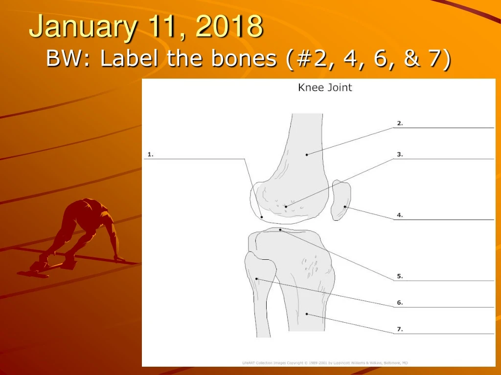
January 11, 2018 BW: Label the bones (#2, 4, 6, & 7)
The Knee Chapter 18
Objectives • Describe the functions of the knee • Describe some of the ligament structure of the knee • Explain the function of the patellofemoral joint • List and define some sports-related injuries of the knee
The Knee • The knee joint is one of the most complex joints in the body • The knee is composed of 3 major bones and muscle groups
Bones of the Knee • The end of the femur flares at its distal end into a pair of rounded prominences called condyles (these are labeled as medial and lateral) • The proximal end of the tibia is flat and is referred to as the tibial plateau • The joint where the femur and tibia come together is called the tibiofemoral joint
Tibiofemoral Joint • Weight-bearing, hinged joint held together with a joint capsule and several important ligaments • The motions at this joint are limited to flexion, extension, and a few degrees of rotation of the tibia on the femur
Cartilage • There are 2 types of cartilage found in the knee joint • The ends of both the tibia and femur are coated with a protective layer of smooth articular cartilage • Provides a smooth surface for gliding of the joint
Cartilage • Between the tibia and femur are 2 crescent-shaped wedges of cartilage called menisci • The medial meniscus: lies between the medial femoral condyle and the medial tibial plateau • The lateral meniscus: lies between the lateral femoral condyle and the lateral tibial plateau
The Menisci • Play several very important roles in the health and function of the tibiofemoral joint • They aid in shock absorption, distribute forces, and improve stability of the femur as it rides on the tibia • They are bathed with synovial fluid
January 22, 2018 • Matching the correct letter to the best description. A. Condyle B. Tibial plateau C. Sesamoid D. Crepitus 1.The rounded prominence found at the point of articulation with another bone 2. A grinding noise or sensation within a joint 3. The top, flat portion of the tibia 4. A small bone formed in a tendon where it passes over a joint **New bell work procedure**
Ligaments of the Knee • There are 4 major ligaments that connect the femur and tibia together • Together they provide stability during walking, running, jumping, or any movement involving the lower extremities
Ligaments of the Knee • Two ligaments are found on the outside of the knee • They are aligned vertically connecting the lateral and medial femur and tibia • These ligaments are called the medial collateral ligament (MCL) and the lateral collateral ligament (LCL)
Ligaments of the Knee • Medial Collateral Ligament (MCL) • Connects the medial femur and the medial tibia • This ligament is in place to help provide stability to the medial knee during activity
Ligaments of the Knee • Lateral Collateral Ligament (LCL) • Connect the lateral femur and the lateral tibia and fibula • Provides stability to the lateral knee during activity
Ligaments of the Knee • There are 2 more ligaments that are located within the knee joint • These ligaments are called the anterior cruciate ligament (ACL) and the posterior cruciate ligament (PCL) • Cruciate comes from the Latin word meaning “cross” • These 2 ligaments cross each other in the knee joint
Ligaments of the Knee • Anterior Cruciate Ligament (ACL) • Attaches to the anterior aspect of the tibial plateau • It crosses the PCL anteriorly and runs from the lateral femur and attaches on the medial tibia • Becoming a fairly common injury in athletics and with the general public
Ligaments of the Knee • Posterior Cruciate Ligament (PCL) • Attaches to the posterior aspect of the lateral tibia • It crosses posteriorly to the ACL and runs from the medial femur and attaches to the lateral tibial plateau
January 24 • Draw a diagram labeling the following knee structures… • Femur • Tibia • Fibula • Patella • ACL,MCL, LCL, PCL
Knee Injuries • Patellofemoral Problems • Not always easy to identify this region as the source of the athlete’s complaints, or to isolate the causative factors from the many possible causes • Understanding the biomechanics of the knee and entire lower extremity is essential for successful management of patellofemoral problems
Knee Injuries • Patellar Tendonitis • Inflammation of the patellar tendon is often seen in sports that involve jumping • Often referred to as jumper’s knee • High force and repetitive strain frequently causes tendonitis of the patellar tendon
Knee Injuries • Medial Collateral Ligament Sprain • Usually caused by a blow to the outside of the knee or a high-energy twisting maneuver • Results in a valgus force on the medial tibiofemoral joint • Pain and tenderness are felt on the medial aspect of the knee • These sprains are categorized by either Grade I, II, III
Knee Injuries • Lateral Collateral Ligament Sprain • Located on the lateral side of the knee and is not frequently involved in sports injuries • Can be injured by a blow to the medial knee which results in a varus stress to the knee joint
Knee Injuries • Anterior Cruciate Ligament Tear: • Usually occurs due to a blow to the posterior knee or a sudden violent twist in the knee during activity • ACL is in place to restrict excessive movement of the tibia during rotation and too much movement of the tibia anteriorly
ACL • ACL tears have become increasingly more common in female athletes over the past couple of decades • Research shows that female athletes who participate in basketball and/or soccer are 4 – 6 times more likely to sustain an ACL injury than males who play the same sport • 70% of ACL injuries in females come from noncontact situations
ACL Tear in Females • Biomechanical Factors: females tend to use their quad muscles more than males. Females tend to land on a flat foot, rather than the toes, which contribute to the increased injury rate • Hormonal Influences: Research is being done to determine if there is an increased chance for tear during menstrual cycle
ACL Tear in Females • Environmental Factors: shoes worn during participation may increase risk of a tear • Anatomical risk factors: the space allowed for the ACL may be smaller than those of male athletes
ACL Tear in Athletes • An example would be a basketball player making a rapid change of direction • Once an ACL is stretched or ruptured, it will not heal • ACL tears are often accompanied by a meniscus and/or MCL sprain • ACL injuries do not follow the same grade scale as MCL or LCL; they are either damaged or not damaged
Posterior Cruciate Ligament Tear • There has not been a lot of research done on PCL injuries because it is injured far less often than other ligaments • Most athletic PCL injuries occur during a fall on the flexed knee with the foot plantar flexed • Another common cause of injury is in motor vehicle accidents
Meniscus Tears • The menisci help make a more concave surface for the condyles to rest and glide on, and thus make the knee joint more stable • The medial meniscus is more easily torn because it is attached more securely than the lateral meniscus • Both menisci can be torn with sudden twist that catches between the femur and tibia and over time due to lack of rubbery consistency
Medial Meniscus Surgery • Stone Clinic :: Videos: Lateral Meniscus Replacement
Osgood-Schlatter Condition • A group of symptoms involving the tibial tubercle epiphysis • The tibial tubercle is a small bump on the tibia where the patella tendon of the quads muscle attaches • This condition effects males ages 12-16 and females ages 10-14
Osgood-Schlatter Condition • The layer under the hard bone of the tibial tubercle is fibrocartilage and is different from any of the body’s other growth cartilage areas • Repetitive traction of the quads may cause disruption and inflammation in the layers of the tubercle • The femur is growing faster than the quads
Define the following: A. Condyle F. Sesamoid B. Valgus G. Synovial fluid C. Tibial plateau H. Effusion D. Patellofemoral joint I. Varus E. Crepitus J.Epiphyseal plate • The medial meniscus lies between? • The menisci are in place to aid in? • The collateral ligaments of the knee are found? • The MCL adds stability during? LCL? • What 2 types of cartilage are found in the knee? • The ACL runs from _________ to __________? PCL? • A MCL sprain often occurs during? • Osgood-Schlatter Condition is a disorder to the? • ACL tears are most often the result from? PCL? • What are some possible reasons why female athletes have a greater incidence of ACL injuries? • Why is Osgood-Schlatter Condition painful?