Download
1 / 1
20 likes | 311 Views
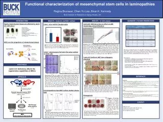
Functional characterization of mesenchymal stem cells in laminopathies. Lamin A/C deficiency affects the regenerative capabilities of MSCs. INTRODUCTION. HYPOTHESIS. Lmna -/- mice exhibit lipodystrophy. RESULTS - in vitro assays. SUMMARY / FUTURE PERSPECTIVES.

E N D
Functional characterization of mesenchymal stem cells in laminopathies Lamin A/C deficiency affects the regenerative capabilities of MSCs INTRODUCTION HYPOTHESIS Lmna-/-mice exhibit lipodystrophy RESULTS - in vitro assays SUMMARY / FUTURE PERSPECTIVES Buck Institute for Research on Aging, Novato, CA REFERENCES CONTACT Regina Brunauer Postdoctoral Research Fellow, Kennedy Lab Buck Institute for Research o n Aging rbrunauer@buckinstitute.org Mainly mesenchymal tissues are affected by lamin A/C deficiency RESULTS – body fat and MSC number Regina Brunauer, Chen-Yu Liao, Brian K. Kennedy APPROACH Introduction Sullivan, T., D. Escalante-Alcalde, et al. (1999). "Loss of A-type lamin expression compromises nuclear envelope integrity leading to muscular dystrophy." J Cell Biol147(5): 913-920. Nikolova, V., C. Leimena, et al. (2004). "Defects in nuclear structure and function promote dilated cardiomyopathy in lamin A/C-deficient mice." The Journal of clinical investigation113(3): 357-369 Li, W., L. Yeo, et al. (2011). "Decreased bone formation and osteopenia in lamin a/c-deficient mice." PloS one6(4). Tong, J., W. Li, et al. (2011). "Lamin A/C deficiency is associated with fat infiltration of muscle and bone." Mechanisms of ageing and development132(11-12): 552-559. Hale, J. S., R. L. Frock, et al. (2010). "Cell-extrinsic defective lymphocyte development in Lmna(-/-) mice." PLoS One5(4): e10127. Results Hu, Y, and Smyth, GK (2009). “ELDA: Extreme limiting dilution analysis for comparing depleted and enriched populations in stem cell and other assays.” Journal of Immunological Methods 347, 70-78. Summary / Future Perspectives Ramos, F., S. Chen, et al. (2012). "Rapamycin reverses elevated mTORC1 signaling in lamin A/C-deficient mice, rescues cardiac and skeletal muscle function, and extends survival." Science translational medicine4(144). Osorio, F., C. Navarro, et al. (2011). "Splicing-directed therapy in a new mouse model of human accelerated aging." Science translational medicine3(106). Lamin A/C deficiency has no effect on MSC morphology and proliferation +/+ -/- • Normal at birth • Growth retardation • Dilated cardiomyopathy • Muscular dystrophy • Lipodystrophy • Osteopenia (Li W et al, PloS One 2011) • Fat infiltration of muscle and bone (Tong J et al, Mech Ageing Dev 2011) • Impaired B- and T cell maturation (Hale JS et al, PloS One 2010) Inguinal fat pad Collagenase digestion Red blood cell lysis Stromal vascular fraction (SVF) (fibroblasts, endothelial cells, preadipocytes, blood cells, MSCs) -/- +/+ +/- Sullivan T et al, J Cell Biol1999 Nikolova V et al, J Clin Invest 2004 Plastic adherence MSCs are the progenitors of mesenchymal tissues Morphology and in vitro proliferation of Lmna-/-MSC. Left: MSC morphology after two weeks in culture. The cells are spindle-shaped and exhibit a characteristic pattern when confluent. No morphologic differences between Lmna+/+(WT) and Lmna-/- (KO) cells are apparent. Scale bar indicates 100 μm. Right:In vitro proliferation. SVF cells were seeded at 5-10x103 cell/cm2. Upon confluence, plastic-adherent cells were split and seeded at 2000 cells/cm2 (passage 1). This was done for all subsequent passages for about 3 months. Cumulative population doublings were calculated and plotted against days in culture. Growth curves were done in duplicates, except for WT C3399. The legend indicates the genotype, the mouse ID number and the replicate number. There may be a small proliferative advantage for Lmna-/- MSCs (KO, red and orange) compared to Lmna+/+ MSCs (WT, blue). Left: Assessment of total body fat by EchoMRI. 5-week-old mice were subjected to live body fat assessment. Lmna-/- mice (KO, red) have about 40% less total body fat compared to Lmna+/+mice (WT, blue). Right: Dissection of the inguinal fat pad for MSC isolation. The fat pads of Lmna-/- mice are considerably smaller. MULTILINEAGE DIFFERENTIATION Multilineage differentiation in vitro SELF-RENEWAL Adipocytes • Most cell lines isolated and tested so far exhibit MSC characteristics such as adipogenic and osteogenic potential, absence of hematopoietic and endothelial surface proteins (CD45, CD31), and presence of stromal surface proteins (Sca1, CD44, CD90, CD105). No apparent differences were found between Lmna-/- and Lmna+/+cell lines. For future analyses, only cell lines exhibiting MSC characteristics will be used. • Next steps are: • Quantification of adipogenic and osteogenic differentiation potential • Chondrogenic and myogenic differentiation • Multilineage differentiation potential of clonogenic MSC populations • Assessment of TOR activity and DNA repair • Characterization of MSC from rapamycin-treated Lmna-/- mice (Ramos F et al, Science Transl Med 2012) • Characterization of MSC from a progeria mouse model (LmnaG609G/G609G) (Osorio FG et al, Science Transl Med 2012) Lmna-/- subcutaneous fat hosts the same number of stem cells Proliferation in vitro Osteoblasts Chondrocytes Lamin A/C-deficient MSC have adipogenic potential MSC CD31-CD45-Sca1+CD90+CD105+ Stroma (supporting hematopoiesis) WT Stem cell number KO 2000 1000 500 100 cells/well Determination of stem cell number in the stromal vascular fraction (SVF) of inguinal fat pads by limiting dilution assay. Following collagenase digestion and red blood cell lysis, SVF cells were seeded in 96 well plates at a density of 100, 500, 1000 and 2000 cells per well. After two weeks, colonies were fixed and stained with crystal violet. The number of wells containing colonies was determined and plotted against the seeded cell number. According to the Poisson single-hit model, one stem cell is present when 37% of the wells contain a colony. The data were analyzed using ELDA, a web-based software (http://bioinf.wehi.edu.au/software/elda/). Left: Example of a crystal violet-stained Lmna+/+(WT) and Lmna-/- (KO) assay plate. Right: Percentage of stem cells in the SVF of Lmna+/+ (WT, blue) and Lmna-/- (KO, red) mouse fat (±95% confidence interval). The x-axis indicates the genotype and the mouse ID number. No gender- or genotype-specific difference has been observed. Adipogenic differentiation of MSC. Cells were seeded in triplicates and grown to confluence. Adipogenesis was induced using MesenCult basal medium with adipogenic supplement (StemCell Technologies; diff). Control cells were treated with growth medium (ctrl). After two weeks, cells were fixed and stained for lipids with oil red O, and nuclei were stained with hematoxylin. All MSC lines tested so far were able to generate adipocytes. Compared to Lmna+/+MSCs (WT), there may be a higher propensity for Lmna-/- MSCs (KO) to differentiate spontaneously in growth medium. Scale bar indicates 100 μm. Cell preparations have MSC surface marker pheno-type Osteogenesis CD90 CD45 Sca1 unstained ctrl Isotypectrl antibody Osteogenic differentiation of MSC. Cells were seeded in triplicates in 24 well plates and grown to confluence. Osteogenesis was induced using StemXVivoosteogenic supplement (RnD systems) in aMEM/10% FBS. Control cells were treated with growth medium. After two weeks, cells were fixed and stained for calcium deposits with alizarin red S. CD31 CD105 CD44 counts No spontaenous differentiation was observed in growth medium. Only wells treated with osteogenic medium are depicted here. Irrespective of the genotype, some MSC lines failed to produce calcified extracellular matrix. A successful second attempt with a subsequent passage of one of these lines (C4419) revealed that this could be due to contamination with other cell types in early passages. fluorescence MSC surface marker phenotype assessed by flow cytometry. Expanded cells were trypsinized, and 2x10⁵ cells/sample were subjected to antibody staining. Data were aquired with a FACSAria instrument, and analyzed with FlowJo. As expected, the isolated cell lines are negative for hematopoietic and endothelial markers (CD45 and CD31) and positive for CD90, CD44, Sca1 and CD105. No differences were observed between Lmna-/- and Lmna+/+cells.