Download
1 / 43
430 likes | 520 Views
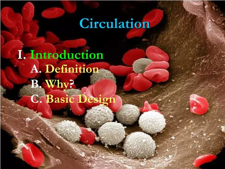
Circulation. I. Introduction. A. Definition. B. Why ?. C. Basic Design. Fluid , vessels , and a pump. Figure 42.6. II. Strategies. A. Gastrovascular Cavity. Cnidarians and Platyhelminthes Thin bodied with one opening Waste is mixed with nutrients. Figure 42.2.

E N D
Circulation I. Introduction A. Definition B. Why? C. Basic Design
Fluid, vessels, and a pump Figure 42.6
II. Strategies A. Gastrovascular Cavity
Cnidarians and Platyhelminthes Thin bodied with one openingWaste is mixed with nutrients Figure 42.2
Echinoderms= Tube feet pull in water to an internal ampulla which communicates with a ring canal to transport water, nutrients, and waste throughout the ring and body. Drainage is out an opening, called the madreporite, on the aboral side.
Arthropods andsome Mollusks fluid, hemolymph, through a muscular pump, sinuses, and ostia back into the tubular hearts. Figure 42.3
Annelids, Mollusks (squids and octopuses), and Vertebratesblood, muscular pump, and vessels, and back to the heart Figure 42.3
III. Vertebrate Design A. Heart 1. Structure a. Strategies
Dependson the habitat and demands Figure 42.4
internal endocardium, middle myocardium, and the external (visceralandparietal = pericardium)epicardium
2. Function a. Valves & Flow
Valves tricuspid & bicuspid andpulmonary & aortic semilunar Papillary muscles and Chordae Tendinae
Valves prevent backflow Figure 42.7
cardiac cycle == systole (contraction) and diastole (relaxation) Figure 42.8
i.Internal input Figure 42.9 ii. External input endocrine system, nervous system, blood pressure, and ion concentrations (Na, Ca, and K)
B. Vessels 1. Structure a. Histology b. Types
Vessels are multi layered (intima, media, and externa (adventia) Figure 42.10
Vessels are multi layered (intima, media, and externa (adventia)
2. Function a. Forces
Blood pressure == force against the wall systole and diastole Figure 42.14 Figure 42.11
Name the forces which can be involved in regulating blood pressure in blood vessels? Blood Volume or Viscosity Cardiac Output Peripheral Resistance Ion Blood Concentrations Hormones
C. Blood 1. Plasma Component
Plasma ==55% of total What is found in the plasma? Figure 42.17
Cellular elements == 45%What cells? Figure 42.15
Blood smear Figure 42.14
White Blood Cell Figure 42.14
Blood Clotting Figure 42.18 Figure 42.14
Blood Clotting Figure 42.14
D. Lymphatic System 1. Components
The lymphatic system == Lymph, vessels, and filters Figure 43.7
Vessels capture lost blood fluid and Figure 43.7
and Nodes filter the lymph to remove material. Figure 43.7