Download
1 / 36
390 likes | 606 Views
The Collapsed Infant (Child) with Suspected Heart Disease (A Pragmatist’s Guide). Nick Pigott Staff Specialist in Paediatric Intensive Care Children’s Hospital at Westmead. PAC 2012, November 16 th and 17 th. Resus room perspective… The collapsed newborn…

E N D
The Collapsed Infant (Child) with Suspected Heart Disease(A Pragmatist’s Guide) Nick Pigott Staff Specialist in Paediatric Intensive Care Children’s Hospital at Westmead PAC 2012, November 16th and 17th
Resus room perspective… • The collapsed newborn… • The case for considering heart disease • Diagnostic approach • Clinical management • (HLHS) • Other customers: • Cardiomyopathy/myocarditis • (arrhythmias) • ‘known’ cardiac patients
Congenital Heart Disease (CHD) accounts for 20% of all congenital malformations
Modes of detection of CHD • Fetal USS • Post natal examination • Six week check • Parental concern/acute presentation
1590 with CHD • (~300,000 live births) • 523 presented before neonatal exam • Routine neonatal examination failed to detect more than half babies with heart disease • Routine exam at 6/52 missed 1/3
Qs Qp ‘Normal’ circulation effectively 2 pumps in series Systemic blood flow (Qs) = Pulmonary blood flow (Qp)
3 main ways babies present with congenital heart disease… • SHOCK(obstructed flow to body) • “BLUE” (obstructed or restricted flow to lungs) • HEART FAILURE(excess volume load, eg. large AV canal defect) [at least the ones intensivists worry about…]
In some newborns with CHD… • Emulating a ‘balanced’ circulation may be dependent on maintaining flow across a persistent arterial duct • Ductal patency can be sustained using an infusion of Prostaglandin E [5 – 100ng/kg/min]
‘Varieties’ of duct-dependent circulation • Duct-dependent mixing • Duct-dependent pulmonary circulation • Duct-dependent systemic circulation
‘Mixing’ lesions Transposition of the Great Arteries (with or without VSD) • Systemic oxygenation dependent on mixing at atrial, ductal levels (+VSD) Presentation:Cyanosis Management: • PGE infusion • May require ventilation for transfer • Definitive balloon atrial septostomy (BAS) • Occasionally babies will have intractable hypoxaemia and metabolic acidosis despite ductal patency – urgent transfer for BAS
Duct-dependent pulmonary circulation(obstructed pulmonary circulation) • Pulmonary atresia with intact ventricular septum • Critical pulmonary stenosis • Tricuspid atresia • Presentation: Cyanosis (murmur) • Management: • PGE infusion • Often results in significant improvement • Aim for percutaneous sats of 70 – 85% • Adequate urine output, absence of metabolic acidosis • May require ventilation for airway control/systemic collapse
Duct-dependent pulmonary circulation Management (continued): • If confident underlying cause is cardiac aim for ‘normo’- ventilation and keep FiO2 between 0.21 and 0.4 HOWEVER! • May be indistinguishable from Persistent Pulmonary Hypertension of the Newborn (PPHN)! • …in which case, will need mechanical ventilation optimised to reduce PVR • Aim for: PaO2 > 80mm Hg (~100% FiO2) pH 7.40 – 7.45 iNO, MgS04,, bicarbonate muscle relaxation, sedation
Duct-dependent systemic circulation(critically obstructed systemic circulation) • Coarctation of the aorta • Interruption of the aortic arch • Hypoplastic left heart syndrome (HLHS) Presentation: Collapse, metabolic acidosis, oliguria, absent femoral pulses NB. Systemic, myocardial (coronary) and pulmonary circulations are all in parallel
Duct-dependent systemic circulation(critically obstructed systemic circulation) • Management: • PGE infusion to re-open duct • AIM is to ‘Balance’ pulmonary, systemic (and coronary) circulations by manipulating PVR and SVR • If collapsed intubation and ventilation • Fluid resuscitation • Inotropes/inodilators (dopamine, adrenaline, milrinone)
Mini summary… • CHD commonest congenital anomaly • Should be considered as underlying cause in presentation of collapsed newborn • Effective clinical mx does not require knowledge of specific underlying lesion
Clinical approach • Physical examination • Cyanosis • Murmur • Pulses • Signs of heart failure (resp rate, hepatomegaly)
Clinical approach… CXR • Heart size • Pul vascularity (too much/too little) ECG
Initial management of the newborn in shock with suspected CHD • Airway Management • IV Access • PGE1 • Antibiotics • Volume, Calcium, Glucose • Supplemental O2 if needed • Crossmatch
Case • Born at 42 weeks gestation. Normal delivery and Apgar score. • Poor feeding and breathlessness noted at 2 days. • Readmitted to hospital. • Hepatomegaly, weak peripheral pulses and acidosis. • Retrieval requested.
Team arrival… • Gasping, grey and grunting • Impalpable peripheral pulses • Single heart sound • CXR: Cardiomegaly • Blood gases: Base deficit of -22, pH = 7.01 • Blood glucose: 1mmol/L • Echo: HLHS, tiny arterial duct
Resuscitation on site • Intubated and ventilated • Paralysed and sedated • Central venous and arterial catheters • Prostaglandin infusion increased • Ventilated • FiO2 = 0.21 (O2 sat = 80%) • pCO2 = 6 • Transferred
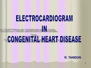
ECG I aVR II aVL III aVF
Parallel circulations • Reduced systemic perfusion = Excessive pulmonary blood flow (PBF) until proven otherwise • Poor Ventricular function = Poor myocardial blood flow = Excessive PBF • Usually, high-dose Inotropes not required
LA RA LV RV HLHS
Other ‘cardiac’ customers… • Cardiomyopathy/myocarditis • (Arrhythmias) • The “known” cardiac patient
Dilated cardiomyopathy (DCM) • 36.5/100 000 • Most commonly within first 2y • Heart usually huge, big liver • Ejection Fraction~10-20%!
Other ‘cardiac’ customers… • Cardiomyopathy/myocarditis • (Arrhythmias) • The “known” cardiac patient
Other ‘cardiac’ customers… • Cardiomyopathy/myocarditis • (Arrhythmias) • The “known” cardiac patient
Summary • Newborns present collapsed as a consequence of congenital heart disease surprisingly often • Consideration of the likely underlying physiology (rather than the exact lesion) may usefully guide clinical management • Some features are more useful than others…
The Money… • Murmur • Cyanosis • Diminished peripheral pulses • Physical signs of heart failure • Prostaglandin infusion • CXR • (ECG) • ?Oxygen might not be the best thing…