Download

1 / 64
650 likes | 679 Views
Explore the structure and function of the skin layers, epidermis, dermis, and subcutaneous layers. Learn about keratinocytes, melanocytes, and skin aging effects. Understand common skin dysfunctions and clinical manifestations, such as psoriasis and allergic contact dermatitis.

E N D
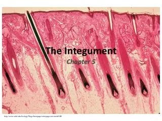
Structure, Function, and Disorders of the Integument Chapter 44
Layers of the Skin • Epidermis • Dermis • Subcutaneous
Layers of the Skin • Epidermis • Stratum basale • Stratum germinativum • Stratum spinosum • Stratum lucidum • Stratum corneum
Layers of the Skin • Epidermis • Keratinocytes • Keratin • Melanocytes • Langerhans cells • Merkel cells
Layers of the Skin • Dermis • Collagen, elastin, reticulum, and a gel-like ground substance • Hair follicles, sebaceous glands, sweat glands, blood vessels, lymphatic vessels, nerves • Fibroblasts, mast cells, macrophages • Subcutaneous layer • Adipocytes • Dermal and subcutaneous collagen are continuous
Layers of the Skin • Dermal appendages • Nails • Hair • Sebaceous glands • Eccrine and apocrine sweat glands • Blood supply • Papillary capillaries
Aging and Skin Integrity • The integumentary system reflects numerous changes from genetic and environmental factors • The skin becomes thinner, drier, wrinkled, and demonstrates a changes in pigmentation • Shortening and decrease in the number of capillary loops • Fewer melanocytes and Langerhans cells • Atrophy of the sebaceous, eccrine, and apocrine glands • Changes in hair color • Fewer hair follicles and growth of thinner hair
Clinical Manifestations of Skin Dysfunction • Macule • Papule • Patch • Plaque • Wheal
Clinical Manifestations of Skin Dysfunction • Nodule • Tumor • Vesicle • Bulla • Pustule
Clinical Manifestations of Skin Dysfunction • Cyst • Telangiectasia • Scale • Lichenification • Keloid • Scar
Clinical Manifestations of Skin Dysfunction • Excoriation • Fissure • Erosion • Ulcer • Atrophy
Clinical Manifestations of Skin Dysfunction • Pressure ulcers • Pressure ulcers result from any unrelieved pressure on the skin, causing underlying tissue damage • Pressure • Shearing forces • Friction • Moisture
Clinical Manifestations of Skin Dysfunction • Pressure ulcers • Stages • Nonblanchable erythema of intact skin • Partial-thickness skin loss involving epidermis or dermis • Full-thickness skin loss involving damage or loss of subcutaneous tissue • Full-thickness skin loss with damage to muscle, bone, or supporting structures
Clinical Manifestations of Skin Dysfunction • Keloids • Elevated, rounded, and firm • Clawlike margins that extend beyond the original site of injury • Excessive collagen formation during dermal connective tissue repair • Common in darkly pigmented skin types and burn scars • Type III collagen is increased.
Clinical Manifestations of Skin Dysfunction • Pruritus • Itching • Most common symptom of primary skin disorders • Itch is carried by specific unmyelinated C-nerve fibers and is triggered by a number of itch mediators • The CNS can modulate the itch response • Pain stimuli at lower intensities can induce itching • Chronic itching can result in infections and scarring due to persistent scratching
Disorders of the Skin • Inflammatory disorders • The most common inflammatory disorder of the skin is dermatitis or eczema • There are various types of dermatitis • The disorders are generally characterized by pruritus, lesions with indistinct borders, and epidermal changes
Inflammatory Disorders • Allergic contact dermatitis • Caused by a hypersensitivity type IV reaction • The allergen comes in contact with the skin, binds to a carrier protein to form a sensitizing antigen; Langerhans cells process the antigen and carry it to T cells, which become sensitized to the antigen • Manifestations • Erythema, swelling, pruritus, vesicular lesions
Inflammatory Disorders • Atopic dermatitis • Type I hypersensitivity—activation of mast cells, eosinophils, T lymphs, and other inflammatory cells • Causes red, weeping crusts and chronic inflammation, lichenification • Irritant contact dermatitis • Nonimmunologic inflammation of the skin • Chemical irritation from acids or prolonged exposure to irritating substances • Symptoms similar to allergic contact dermatitis • Treatment—remove stimulus
Inflammatory Disorders • Stasis dermatitis • Occurs in the legs as a result of venous stasis, edema, and vascular trauma • Sequence of events: erythema, pruritus, scaling, petechiae, ulcerations • Seborrheic (sebōrēik) dermatitis • Inflammation of the skin involving the scalp, eyebrows, eyelids, nasolabial folds, and ear canals • Scaly, white, or yellowish plaques
Papulosquamous Disorders • Psoriasis • Chronic, relapsing, proliferative skin disorder • T cell immune–mediated skin disease • Scaly, thick, silvery, elevated lesions, usually on the scalp, elbows, or knees caused by a high rate of mitosis in the basale layer • Shows evidence of dermal and epidermal thickening • Epidermal turnover goes from 26-30 days to 3-4 days • Cells do not have time to mature or adequately keratinize
Papulosquamous Disorders • Psoriasis • Plaque psoriasis • Inverse psoriasis • Guttate psoriasis • Pustular psoriasis • Erythrodermic psoriasis
Papulosquamous Disorders • Pityriasis rosea • Benign, self-limiting inflammatory disorder • Usually occurs during the winter months • Herald patch • Circular, demarcated, salmon-pink, 3- to 4-cm lesion
Papulosquamous Disorders • Lichen planus • Benign, inflammatory disorder of the skin and mucous membranes • Unknown origin, but T cells, adhesion molecules, inflammatory cytokines, and antigen presenting cells are involved • Nonscaling, violet-colored, 2- to 4-mm lesions • Wrists, ankles, lower legs, genitalia
Papulosquamous Disorders • Acne vulgaris • Inflammatory disease of the pilosebaceous follicles • Acne rosacea • Inflammation of the skin that develops in adulthood • Lesions • Erythematotelangiectatic, papulopustular, phymatous, and ocular • Associated with chronic, inappropriate vasodilation resulting in flushing and sensitivity to the sun
Papulosquamous Disorders • Lupus erythematosus • Inflammatory, autoimmune disease with cutaneous manifestations • Discoid lupus erythematosus • Restricted to the skin • Photosensitivity • Butterfly pattern over the nose and cheeks • Systemic lupus erythematosus
Vesiculobullous Disorders • Pemphigus • Rare, chronic, blister-forming disease of the skin and oral mucous membranes • Blisters form in the deep or superficial epidermis • Autoimmune disease caused by circulating IgG autoantibodies • The antibodies are against the cell surface adhesion molecule, desmoglein in the suprabasal layer of the epidermis
Vesiculobullous Disorders • Pemphigus • Tissue biopsies demonstrate autoantibody presence • Types • Pemphigus vulgaris (severe) • Pemphigus foliaceus • Pemphigus erythematosus
Vesiculobullous Disorders • Bullous pemphigoid • More benign disease than pemphigus vulgaris • Bound IgG and blistering of the subepidermal skin layer • Subepidermal blistering and eosinophils distinguish pemphigoid from pemphigus
Vesiculobullous Disorders • Erythema multiforme • Acute, recurring disorder of the skin and mucous membranes • Associated with allergic or toxic reactions to drugs or microorganisms • Caused by immune complexes formed and deposited around dermal blood vessels, basement membranes, and keratinocytes • “Bull’s-eye” or target lesion • Erythematous regions surrounded by rings of alternating edema and inflammation
Vesiculobullous Disorders • Erythema multiforme • Bullous lesions form erosions and crusts when they rupture • Affects the mouth, air passages, esophagus, urethra, and conjunctiva • Severe forms • Stevens-Johnson syndrome (bullous form) • Toxic epidermal necrolysis
Infections • Bacterial infections • Folliculitis • Furuncles • Carbuncles • Cellulitis • Erysipelas • Impetigo
Infections • Viral infections • Herpes simplex virus • Herpes zoster and varicella
Warts • Benign lesions caused by the human papillomavirus (HPV) • Diagnosed by visualization • Condylomata acuminata • Venereal warts
Fungal Infections • Fungi causing superficial skin lesions are called dermatophytes • Fungal disorders are called mycoses; mycoses caused by dermatophytes are termed tinea • Tinea capitis (scalp) • Tinea pedis (athlete’s foot) • Tinea corporis (ringworm) • Tinea cruris (groin, jock itch) • Tinea unguium (nails) or onychomycosis
Fungal Infections • Candidiasis • Caused by Candida albicans • Normally found on the skin, in the GI tract, and in the vagina • C. albicans can change from a commensal organism to a pathogen • Local environment of moisture and warmth, systemic administration of antibiotics, pregnancy, diabetes mellitus, Cushing disease, debilitated states, age younger than 6 months, immunosuppression, and neoplastic diseases