Download
1 / 56
630 likes | 1.06k Views
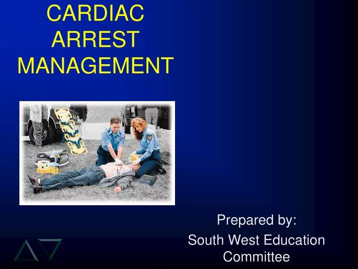
CARDIAC ARREST MANAGEMENT. Prepared by: South West Education Committee. SWEC MEMBERS. Cambridge – Lori Smith Grey Bruce – Andy Whittemore Hamilton – Ken Stuebing, Tim Dodd Lambton – Judy Potter London – Tre Rodriguez Niagara – Greg Soto Windsor – Cathie Hedges RTN – Peter Deryk.

E N D
CARDIAC ARRESTMANAGEMENT Prepared by: South West Education Committee
SWEC MEMBERS • Cambridge – Lori Smith • Grey Bruce – Andy Whittemore • Hamilton – Ken Stuebing, Tim Dodd • Lambton – Judy Potter • London – Tre Rodriguez • Niagara – Greg Soto • Windsor – Cathie Hedges • RTN – Peter Deryk
“The Power of 7” Base Hospital Programs • Goal: One single certification for all of SouthWestern Ontario by Fall 2005!! • Recert process same across SW this year. • Notice, all paperwork will say SWEC. • Some information may not be specific to Hamilton BH or Services in our area. • Pictures for data base in one of the stations
COURSE OVERVIEW • Chain of Survival • Review of the conduction system • Cardiac Monitoring • Protocols • Special circumstances • CPR & SAED reminders
CHAIN OF SURVIVAL • Early Access (911) • Someone must realize there is an emergency and act quickly to initiate the EMS. • Early CPR • A trained individual starts CPR at once to help maintain a viable heart until help arrives. • Early Defibrillation • First responder arrives with the training and equipment to defibrillate the heart. As time increases chances for survival decrease. • Early Advanced Life Support • ALS within minutes increases the chance of survival.
CAUSES OF CARDIAC ARREST • # 1 Cause = Conduction Disturbances • # 2 Cause = AMI / ischemia Other Causes include: • Traumatic • Hypoxia / Respiratory • Metabolic
RHYTHM INTERPRETATION • 5 Steps Approach • Step 1: What is the rate? • brady < 60 bpm, tachy > 100 bpm • Step 2: Is the rhythm regular or irregular? • Step 3: Is there a P wave - is it normal? • are P waves associated with each QRS? • Step 4: P-R Interval/relationship? • PR interval (normal 0.12 - 0.20 sec) • Step 5: Normal QRS complex? • Normal QRS complex < 0.12sec
LETHAL DYSRHYTHMIAS • There are four major life threatening Pulseless Dysrhythmias: • NON SHOCKABLE RHYTHMS 1) Asystole - Flat Line 2) PEA - Pulseless Electrical Activity • SHOCKABLE RHYTHMS 3) VF - Ventricular Fibrillation 4) VT - Pulseless Ventricular Tachycardia
Asystole • No heart electrical activity • No excitation of the heart muscle • No Cardiac output • Usually the terminal rhythm of a an unsuccessful cardiac resuscitation
Normal Sinus Rhythm • Usually represented by a normal functioning electrical conduction system • Heart Rate average is 72 beats / minute
Pulseless Electrical Activity • A rhythm is determined to be PEA when your pulseless patient presents with a rhythm which you would normally expect to produce some form of cardiac output. • DO NOT assume that since there is a rhythm on the screen that the patient has a pulse!!
Ventricular Tachycardia • Stimulus is originating from the ventricles • Loss of atrial kick may lead to Inadequate ventricular filling couple with the increased rate causes: • Poor cardiac output, may or may not produce a pulse • Most SAED units will only shock if heart-rate is > 180 B.P.M.
Ventricular Fibrillation • No organized excitation of heart muscle • Heart is physically quivering compared to contracting (seizing) • No Cardiac Output
Defibrillation and Time • Approximately 50% survival after 5 minutes • Survival reduced by 7% to 10% per minute (with no CPR) • Rapid defibrillation is key • CPR prolongs VF, slows deterioration Minutes: collapse to 1st shock
Defibrillation • Defibrillation applies electrical energy to the heart muscle. • This energy causes depolarization of all heart cells at the same time. • Therefore all repolarize at the same time. • We hope this starts an organized perfusing rhythm • We only apply a shock, via the S.A.E.D, to the heart of a VSA patient
Step 1: Rate? • Step 2: Regular or irregular? • Step 3: Is the P wave normal? • Step 4: P-R Interval/relationship? • Step 5: QRS complex < 0.12 sec? ~ 90 bpm Irregular P waves normal, extra beats have associated P wave 0.12 - 0.20 sec Yes PACs
Step 1: Rate? • Step 2: Regular or irregular? • Step 3: Is the P wave normal? • Step 4: P-R Interval/relationship? • Step 5: QRS complex < 0.12 sec? Variable < 100 Irregularly Irregular No P waves None Yes Atrial Fibrillation
Step 1: Rate? • Step 2: Regular or irregular? • Step 3: Is the P wave normal? • Step 4: P-R Interval/relationship? • Step 5: QRS complex < 0.12 sec? Variable ~ 100 Irregular P waves Associated with most QRS Yes - not all Yes - not all PVC - unifocal
Step 1: Rate? • Step 2: Regular or irregular? • Step 3: Is the P wave normal? • Step 4: P-R Interval/relationship? • Step 5: QRS complex < 0.12 sec? 150 Regular No P waves N/A Yes Accelerated Juntional
Step 1: Rate? • Step 2: Regular or irregular? • Step 3: Is the P wave normal? • Step 4: P-R Interval/relationship? • Step 5: QRS complex < 0.12 sec? 40-70 Irregular P waves regular Not always with a QRS longer each beat Yes Second Degree AV Block Type 1
Step 1: Rate? • Step 2: Regular or irregular? • Step 3: Is the P wave normal? • Step 4: P-R Interval/relationship? • Step 5: QRS complex < 0.12 sec? < 30 bpm Regular P waves normal, not with QRS None Yes 3rd degree Heart Block
TAKE HOME POINTS • Use the 5 step approach. • Remember where the lead is and what it should look like. (lead placement can effect what you see) • Use it or lose it. • Remember normal electrical conduction path and rates. • The monitor is a voltage gauge not a pressure gauge - check the Pulse!
GUIDELINES • 10 second pause between shock and subsequent analysis to prevent accidentally missing a shockable rhythm ASYSTOLE for 6 SEC V FIB V FIB SHOCK DELIVERED
If Protocol ends with 3 “No Shocks” in a row • If you receive: • 3 “Check Patient” messages in a • 2 minute time frame • STOP the vehicle and Analyze • Result in: • 1 no shock • 1 stack of 3 shocks 3 2 1 GO
DEFIBRILLATOR ERRORS • If the defibrillator fails during a call, complete the following actions. • Check the adherence of the pads;change pads if required • Check the cables and connections • Change the battery • ALL these actions should take no longer than 60seconds • If you cannot solve the problem, abandon the protocol and continue with BCLS only
When is the Defibrillator not attached to a VSA patient? • Age < 8 years old • Penetrating trauma • Obviously Dead
Criteria for Obviously Dead • Physical Findings: • VSA • Decapitation • Transection • Decomposition (Consider time frame of arrest) • lividity / mottling / putrefaction • Gross rigor mortis • Gross Charring • Gross cranial or visceral contents.
SPECIAL SITUATIONS • Vomiting patient during charge up • Pacemakers • Automatic Implantable Cardioverter Defibrillator(AICD) • DNR orders • unless the patient falls under the MOH Interfacility DNR directive, DNR orders will NOT be recognised in the field
SPECIAL SITUATIONS Pacemaker or AICD • Avoid placing pads directly over. • Apply pads at least 1 to 2 inches away. • Follow all protocols.
SPECIAL SITUATIONSWet patient • Victim lying in water. • Once on land, dry patient before applying SAED. • Remember, let the rescue experts do the rescuing.
SPECIAL SITUATIONS Medication patches • Transdermal medication patches: blocking pad placement? • While wearing gloves, remove patch and wipe area with alcohol wipe and dry. • Place AED pads and follow protocol.
SPECIAL SITUATIONS Paediatric Arrest • Age: victim <8 years old? • CPR only.
SPECIAL SITUATIONS Hypothermia • Hypothermia • Definition: core body temperature <35°C • Causes: exposure to extreme cold ( damp)
HYPOTHERMIA Clinical Signs and Symptoms • Lethargystuporcoma • Muscle rigidity, cessation of shivering • Dilated pupils, nonreactive pupils • bradycardia, slow AF, VF, or asystole
HYPOTHERMIA Initial Therapy • Remove wet garments • Protect against heat loss and wind chill (use blankets and insulating equipment) • Maintain horizontal position • Avoid rough movement and excess activity • Gradually re-warm • High flow oxygen via NRB • Monitor cardiac rhythm
1 NO SHOCK ANYWHERE Check pulse No Pulse CPR concurrent with transport 3 SHOCKS TOTAL Shock #1 Shock #2 Shock #3 Check Pulse No Pulse CPR transport HYPOTHERMIACardiac Arrest
HYPOTHERMIAGeneral Approach • Maintain horizontal position • Vertical position may compromise cerebral and systemic perfusion • Avoid rough movements and activities • Handle victim gently during CPR, BVM ventilation and transport
SPECIAL SITUATIONSTraumatic Cardiac Arrest • This protocol does not include VSA patients as a result of penetrating trauma. • After adequate airway and c-spine management, apply AED and proceed with the following algorithm if Blunt Trauma is the suspected cause of the arrest.
1 NO SHOCK ANYWHERE Check pulse No Pulse CPR concurrent with BTLS care Transport 3 SHOCKS TOTAL Shock #1 Shock #2 Shock #3 Check pulse No Pulse CPR concurrent with BTLS care Transport Blunt Trauma Protocol
Traumatic Cardiac Arrest • If cardiac arrest is caused by penetrating trauma • Package the patient and transport immediately without initiating SAED protocols.
1 NO SHOCK ANYWHERE Check pulse No Pulse CPR Transport 3 SHOCKS TOTAL Shock #1 Shock #2 Shock #3 Check pulse No Pulse CPR Transport Airway Obstruction Ventilate - Reposition - Ventilate Perform visualisation of airway q 15 compressions If cleared start protocol minus shocks delivered