BME 101 Biomedical Optics and Lasers
BME 101 Biomedical Optics and Lasers. Instructor: Irene Georgakoudi TA: Cherry Greiner Class meeting time: T/Th: 10:30 -11:45 AM Office hours: Tuesdays, 3:00-4:30 PM Blackboard site: http://blackboard.tufts.edu Reading material on reserve (Tisch):

BME 101 Biomedical Optics and Lasers
E N D
Presentation Transcript
BME 101 Biomedical Optics and Lasers Instructor: Irene Georgakoudi TA: Cherry Greiner Class meeting time: T/Th: 10:30 -11:45 AM Office hours: Tuesdays, 3:00-4:30 PM Blackboard site: http://blackboard.tufts.edu Reading material on reserve (Tisch): Handbook of Biomedical Photonics, Tuan Vo Dinh Introduction to Biophotonics, Prasad Biological spectroscopy, Campbell Electo-optics library: Optics, Hecht
Evaluation • 10% Class participation • 30% Homeworks/Lab reports (due every Tuesday) • 15% Midterm exam • 25% Final exam • 20% Final paper and presentation • 10-15 page paper on specific molecular imaging method and its impact on a clinical problem • Introduction • Theoretical background of method • Background on clinical problem • Instrumentation • Methods/Results • Advantages/Disadvantages • Suggested improvements
What is biomedical optics? • Biomedical optics is typically defined as the area of study of methods/technologies based on the use of visible light (applications cover UV-NIR) for: • Improving basic understanding of biological processes (from gene to tissue level) • Enhancing the detection and treatment of human diseases (from acne to atherosclerosis and cancer)
Syllabus • Basic principles • Spectroscopic methods • Microscopy and Imaging • Photodynamic Therapy/Flow Cytometry
Basic Principles • Light matter interactions • Basic wave definitions • Schrodinger’s equation -Bonds and orbitals -Biological chromophores
Basic Principles • Laser basics • Principles of operation • Stimulated emission • Critical inversion • Pumping schemes • Major laser components • Laser beam properties • Diode lasers
Cell and Tissue basics • Cell basics • Major cellular components • Origins of intrinsic cellular optical signals • Tissue basics • Major epithelial types • Connections to disease and optical sources of contrast
Spectroscopic methods • Absorption • Scattering • Fluorescence
Adenoma 20 18 16 14 Normal x (cm) 12 10 8 6 4 2 0 0 2 4 6 8 10 y (cm) Spectroscopic methods • Basic Theoretical principles • Instrumentation • Applications Breast cancer atherosclerosis Colon cancer
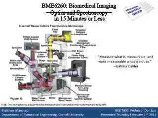
Microscopy and Imaging • Basic Principles
Confocal in vivo H&E “En face” SECTION of human skin From animals to humans From organelles to cells Tumors and blood vessels imaged in vivo From cells to tissues From tissues to animals 4-Pi microscopy Mitochondrial network of live bacterial cell 80 nm res Triple stained endothelial Cell of pulmonary artery Engineered tissue: Fibroblast (red) in collagen matrix (green) Endogenous signal
Optical imaging of cell-matrix interactions Collagen gel embedded with GFP-expressing fibroblasts Leica TCS SP2 confocal microscope Excitation wavelength: 488 nm Green channel: 485-490 nm (scattering) Red channel: 500-620 nm (GFP fluorescence) Stack size: 238x238x125 mm Images acquired using 63X, 1.2 NA, water immersion objective
Photodynamic Therapy • Basic Principles • Applications Macular degeneration
Flow Cytometry • Basic Instrumentations • Advanced Methods
Reading assignment: • Introduction to biophotonics, ch. 1 and 2.1 • Lecture notes • Also posted on the blackboard site: • Calculus review • Electromagnetic waves review
Biomedical optics Exploiting interactions of light with matter Wavelengths used typically: 300-900 nm
Why biomedical optics? • Major advantages: non-invasive; high resolution • Continuous or repetitive monitoring • Study/characterize process/disease in natural environment (no artifacts) • More sensitive/accurate monitoring • Real-time information • Triage with therapy • Accurate dosimetry • Psychological impact
Plato (427-347 BC) Believer of extramission theory: Eye emits a “fire” providing man the capability of vision by seizing objects It all started with the Greeks…
Aristotle (384-322 BC) Light emitted by a source is captured by the eyes when reflected by an object It all started with the Greeks…
Euclid (circa 325-265 BC) Treatise entitled “Catoptrics” Foundations of geometric optics First law of reflection It all started with the Greeks…
Galen of Pergamum (Claudius Gelenus: 130-201 AD) Described anatomical details of the eye Identified lens as principle eye instrument Believed in extramission theory It all started with the Greeks…
Mohammad ibn Zakariya al-Razi (864-930AD) Also known as Rhazes Observed that pupil contracts in response to light Philosophers from the Middle-East followed…
Abu Ali al-Hasan ibn al-Haytham (965-1040 AD) Also known as Alhazen Considered by some as the father of Optics Wrote comprehensive treatise on optics (Katib-al-Manazir/Book on Optics), translated in Latin in 1270 Proved that extramission theory is not correct Detailed description of human eye Theory of vision which prevailed until 17th century Discussed primary and secondary light sources, light propagation and colors Studied spherical and parabolic mirrors Laws of reflection and refraction Philosophers from the Middle-East followed…
Leonardo da Vinci (1452-1519 AD) Initially believed in extramission, but later changed his view in support of external light sources based on experiments he performed with ox eyes Western philosophers/scientists
Johannes Kepler (1571-1630) Established retinal image formation theory based on experiments with ox eyes Law of refraction for small angles of incidence Western philosophers/scientists
Only corpuscular theory of light prevalent until 1660 Francesco Maria Grimaldi (Bologna) described diffraction in 1660 Theories on nature of light:Light as a particle vs. Light as a wave
Sir Isaac Newton (1642-1727) Embraces corpuscular theory of light because of inability to explain rectilinear propagation in terms of waves Demonstrates that white light is mixture of a range of independent colors Different colors excite ether into characteristic vibrations---sensation of red corresponds to longer ether vibration Light as a particle
Christiaan Huygens (1629-1695) Huygens’ principle (Traite de la Lumière, 1678): Every point on a primary wavefront serves as the source of secondary spherical wavelets, such that the primary wavefront at some later time is the envelope of these wavelets. Wavelets advance with speed and frequency of primary wave at each point in space Light as a wave http://id.mind.net/~zona/mstm/physics/waves/propagation/huygens1.html
Thomas Young (1773-1829) 1801-1803: double slit experiment, showing interference by light from a single source passing though two thin closely spaced slits projected on a screen far away from the slits Light as a wave http://vsg.quasihome.com/interfer.htm
Augustine Fresnel (1788-1827) 1818: Developed mathematical wave theory combining concepts from Huygens’ wave propagation and wave interference to describe diffraction effects from slits and small apertures Light as a wave
Michael Faraday (1791-1865) 1845: demonstrated electromagnetic nature of light by showing that you can change the polarization direction of light using a strong magnetic field Electromagnetic wave nature of light
James Clerk Maxwell (1831-1879) 1873: Theory for electromagnetic wave propagation Light is an electromagnetic disturbance in the form of waves propagated through the ether Electromagnetic theory
1900: Max Planck postulates that oscillating electric system imparts its energy to the EM field in quanta 1905: Einstein-photoelectric effect Light consists of individual energy quanta, photons, that interact with electrons like particle 1900-1930 it becomes obvious that concepts of wave and particle must merge in submicroscopic domain Photons, protons, electrons, neutrons have both particle and wave manifestations Particle with momentum p has associated wavelength given by p=h/l QM treats the manner in which light is absorbed and emitted by atoms Quantum mechanics Max Planck Heisenberg Niels Bohr Schrödinger Louis de Broglie
Classical Description of Light Wave Equation (derived from Maxwell’s equations) Any function that satisfies this eqn is a wave It describes light propagation in free space and in time (see calculus review handout)
Classical Description of Light Plane Wave Solution One useful solution is for plane wave E r B
Classical description of light Considering only the real part of the previous solution to make things simpler, we have for the electric field propagating along one dimension, z (or distance)
Period time
Light as a wave: Basic concepts Phase=f=wt-kz • Phase of a wave is the offset of the wave from a reference point fo • We typically talk about a phase shift • When light interacts with matter (e.g. as it travels through a biological specimen), its speed of propagation slows down. The wave emanating from the specimen exhibits a phase shift when compared with the initial wave • The refractive index , n, and the thickness of a specimen determine by how much the wave is retarded Green-incident wave Blue-wave after passing through specimen shifted by l/4
Phase=f=wt-kz Coherent Light Monochromatic (only one wavelength/frequency) waves traveling in phase Incoherent Light Monochromatic (only one wavelength/frequency) waves traveling out of phase Incoherent Light
Constructive interference Destructive interference