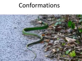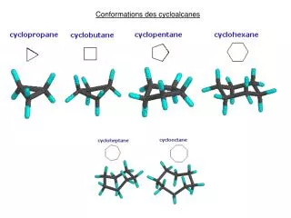
Retinal Conformations
Retinal Conformations. SRJC May 12 th 2008 Younes Ataiiyan Physics 43. Timothy Montague. Nazareth Tesfai. University of Rochester, Institute of Optics. Table 1.0 – Retinal forms. Biologically, retinal (a form of vitamin A) is stored in the

Retinal Conformations
E N D
Presentation Transcript
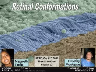
Retinal Conformations SRJC May 12th 2008 Younes Ataiiyan Physics 43 Timothy Montague Nazareth Tesfai University of Rochester, Institute of Optics
Table 1.0 – Retinal forms Biologically, retinal (a form of vitamin A) is stored in the membranes of the rods and cones of optical organs (e.g. the eye). 11-Cis-Retinal is the primary visual pigment for all animals, while some animals have additional visual pigments as listed in Table 1.0. Source: Nakanishi 1991 2 µm in diameter 45 µm in length photon Rod Cell
11-Cis-Retinal Beta-carotene Where does 11-cis retinal come from? Beta-carotene as an optical material in non-linear nano-circuits. Molar Mass: 536.873 g/mol In the small intestine (mucosa), beta-carotene (above) is cleaved in half by an enzyme called beta-carotene dioxygenase to produce 11-Cis-Retinal. Beta-carotene is found in carrots,mangos,etc. Molar Mass: 284.436g/mol Floating in the lipid membranes discs of the rod and cone cells are proteins known as opsin (GMW ≈ 4000 g/mol). Opsin holds the 11-cis-retinal molecule between seven alpha helices.
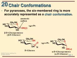
Carbon-Carbon double bond (CIS: Hydrogen's on same side) Source: Wikipedia H H H H • 11-cis retinal • 11-trans retinal B Carbon-Carbon double bond (TRANS: Hydrogen's on opposite side) 11-cis retinal is a non-polar molecule; like beta-carotene it is in the molecular family known as the Cartenoids. PHOTOISOMERIZATION: When a photon of blue light (λ = 440-490 nm) strikes the retinal molecule it breaks an alkene bond (C=C) by exciting an electron. This allows the 11-cis retinal conformation to isomerize into 11-trans retinal (which is more stable, lower overall molecular energy).
EXCITING AN ELECTRON: When a photon excites an electron in a p-orbital, It jumps from the ground state (zero potential energy) at π into a higher potential energy state at π*. As identified by the infinite potential well model. V= V= Infinite Potential Well x=0 x=L V=0
Breaks double bond into a single A photon excites a π electron Reformation of Double bond Rotation about single bond Carbon Carbon n=2 n=1 11-trans retinal 11-cis retinal S = sharp P = principal MO Diagram Steric Hindrence
HOW DOES THIS CONFORMATION AFFECT VISION? MONOCHROMATIC VISION: Photoisomerization of 11-cis retinal into 11-trans retinal, induces a conformational change in opsin that triggers a second messenger cascade – ultimately responsible for monochromatic vision in the dark. COLOR VISION: Closely related opsins which differ only in a few amino acids (and absorbed wavelengths) are responsible for color vision.
Conclusion Optical organs are comprised of rods and cones, which in turn house rhodopsin (opsin + retinal). 11-Cis retinal (cofactor) undergoes photoisomerization, to 11-trans retinal. More specifically; when an electron in a p-orbital (of the π bond) is excited by a photon (λ = 440-490 nm), the electron jumps (by infinite well model) into an anti-bonding orbital π* (higher potential energy) - which breaks the π bond. 11-cis retinal then rotates around the σ bond (C-C) due to steric hinderence of the methyl substituents, and to a trans configuration. The molecule is now in a lower molecular energy conformation. When the electron returns to the ground state (zero potential energy) at π, the double bond reforms (C=C) and the molecule is called 11-trans retinal. 11-trans retinal is longer in length then 11-cis retinal which expands the circumference of the seven opsin alpha-helices, and triggers a second massager cascade - which ultimately is responsible for the perception of monochromatic light.
Works Cited Websites: http://www.diginfo.tv/archives/2006/02/09/national_institute_of_advanced_22.html#more www.ks.uiuc.edu/Research/rhodopsin/ http://webexhibits.org/colorart/ www.circadian.org/biorhyt.html http://www.accessexcellence.org/AE/AEC/CC/vision_background.php http://www.optics.rochester.edu/workgroups/cml/opt307/spr06/joe/index.htm http://en.wikipedia.org/wiki/Retinal http://en.wikipedia.org/wiki/Rhodopsin Images: http://www.hortcouncil.ca/images/carrots.jpg http://upload.wikimedia.org/wikipedia/commons/archive/d/da/20060520170505!Beta-carotene.png http://education.vetmed.vt.edu/Curriculum/VM8054/EYE/ROD.HTM http://www.dark-layouts.net/Backgrounds/purple/images/purple_nebula.jpg