Download
1 / 83
830 likes | 997 Views
Somatic & Special Senses. Types of Sensory Receptors. Chemoreceptor - Stimulated by changes in chemical concentrations of substances Pain receptor -stimulated by damage to tissue. Types of Receptors Cont’d. 3. Thermoreceptor -stimulated by changes in temperature 4. Mechanoreceptor

E N D
Types of Sensory Receptors • Chemoreceptor -Stimulated by changes in chemical concentrations of substances • Pain receptor -stimulated by damage to tissue
Types of Receptors Cont’d 3. Thermoreceptor -stimulated by changes in temperature 4. Mechanoreceptor - Stimulated by changes in pressure or movement 5. Photoreceptor - Stimulated by light energy
Why are you not aware of every sensory input every minute? • As you sit in this classroom, why are you not always 100% aware of the clock ticking or the air conditioner running? • Sensory Adaptation- brains ability to prioritize sensory input it receives and ignore unimportant stimuli.
Sensations • Definition: A feeling that occurs when the brain interprets sensory impulses. • Projection: the brain causes a sensation to seem to come from the region of the body being stimulated.
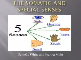
Somatic Senses • Somatic Senses are associated with receptors in the skin. • Out of the 5 types of receptors, which ones do you think could be stimulated by the skin? • Mechanoreceptor • Thermoreceptor • Pain Receptor
Mechanoreceptors- Touch & Pressure Senses • Free nerve endings • Meissner’s corpuscles- found mostly in hairless parts of skin (ex: lips, fingertips, palms, soles). Respond to light touch. • Pacinian corpuscles- found in deeper parts of tissue (ex: tendons & ligaments). Respond to heavy pressure.
Temperature Senses • Warm receptors - Most receptive between 77°F to 113°F - Above 113°F stimulate pain receptors burning sensation • Cold receptors - Most sensitive between 50°F to 68°F - Below 50°F stimulate pain receptors freezing sensation ** Both receptors adapt quickly. This is why after a while your feeling of warmth or cold will begin to fade.
Pain Receptors • Protect the body because damaged tissues stimulate them. • Pain receptors adapt poorly
What is visceral pain? • Pain that may feel as if it is coming from some part of the body other than the part being stimulated. • Examples??? • Heart: feel pain in left arm • Why does this happen? • Because pain impulses of the heart travel over the same nerve pathways of the skin of the left arm. Brain (cerebrum) may misinterpret where the source of pain comes from.
Pain Nerve Fibers • Acute pain fibers- thin, myelinated. Rapid impulses sent shooting pain • Chronic pain fibers- thin, unmyelinated. Slower impulses sent dull, aching sensation
Regulation of Pain Impulses • Thalamus- awareness of pain • Cerebral Cortex- determine pain intensity, pain source, emotional & motor responses to pain. • Enkephalins & endorphins are released in response to extreme pain & provide natural pain control. • How? • Bind to neuron receptors/channels…slow down/stop nerve impulses from passing through
Sense of Smell • Olfactory (smell) receptors are chemoreceptors • Chemicals dissolved in liquids stimulates it. • In the nasal passage- lots of cilia (hairlike projections) come off of olfactory receptor cells
How it works • Odorant molecules bind to the cilia in different patterns stimulates cell impulse sent to the brain. • Combinational olfactory code • Example: if there are 10 odor receptors, chocolate might be receptor 6,7,10 while garlic is 1,3,5
Why do you have to smell deeply for faint scents? • Olfactory organs & receptors are high in the nasal cavity above where usual inhaled air passes.
Comparing Humans to Animals • 60% of genes that encode for human olfactory receptor proteins have mutated to be inactive. • Other animals (mice, monkeys, dogs, etc.) have a higher % of genes encoded to be active • Why could this be? • Humans do not rely on their sense of smell to find food as much as other animals. • # of olfactory receptor cells… • Human: 12 million • Bloodhound: 4 billion
Taste • How many taste buds do we have on the surface of our tongue? • 10,000 • On the roof & walls of throat? • 1,000 • Tiny elevations are called papillae
Taste Receptors • Each taste bud has 50-150 receptor cells, each replaced every 3 days. • Taste pore has taste hairs that protrude from the cells. • Sensory nerves are next to the receptor cells and get stimulated by receptor cells.
Why is saliva important? • Before a particular chemical can be tasted, it must be dissolved in the watery fluid surrounding the taste buds (saliva) • Salivary glands secrete saliva.
Taste (Gustatory) Sensations • Four primary sensations • Sweet • Sour • Salty • Bitter **What is the difference between sour & bitter? -sour: lemon -bitter: caffeine
Tongue Mapping • The taste buds on the tongue are, of course, important for the flavor of food. See if different parts of the tongue are most sensitive to different characteristics of food (i.e., salty, bitter, sour, sweet). Get examples of each of these tastes (for example, salty water, sugary water, vinegar for sour and coffee for bitter). • PROCEDURE: Dip the toothpicks into the solutions and lightly touch the tongue. Repeat the tests on different portions of the tongue. It may help to drink a bit of water in between tests. Also be careful in testing the back part of the tongue...some people may gag! Are parts of the tongue more sensitive to specific flavors or are all parts of the tongue equally sensitive to the flavors? If so, indicate on a drawing of the tongue the areas that are most sensitive to the different tastes. Compare tongue drawings with tongue drawings from other people.
Where are each of these sensations predominate on the tongue?
What is that flavor?? • Flavors result from a combination of primary sensations. • Also involves smelling & feeling the texture & temperature of foods. • Some foods may also stimulate pain receptors to give a burning sensation
Relationship Between Smell & Taste • Both chemoreceptors • Function closely together & aid in food selection • Both smell & taste adapt rapidly • This is why you do not lose the taste of something as you eat it b/c it stimulates different receptors on your tongue
Eye Structure • Visual Accessory Organs • Assist the eye in providing vision • Eyelids & Lacrimal apparatus • Protect the eye • Extrinsic muscles • Move the eye
The Eyelid • Has 4 layers • Skin • Thinnest skin of the body • Muscle • Open & close eye • Connective Tissue • Conjunctiva • Mucous membrane that lines the inner surface of the eyelids and folds back to cover the anterior surface of the eyeball, except for the cornea.
Lacrimal Apparatus • Lacrimal Gland: Secretes tears • Series of ducts: carry tears to nasal cavity • Tears are secreted continuously to lubricate the surface of the eye and lining of the lid. • Tears drain into nasal cavity • Tears also contain an enzyme, Lysozyme, which reduced the risk of eye infection.
Extrinsic Muscles • 6 types which rotate the eye in various directions. 1. Lateral rectus 2. Medial rectus 3. Inferior rectus 4. Superior rectus 5. Inferior oblique 6. Superior oblique
Eye Structure • Hollow, spherical shape • 2.5 cm in diameter • Has 3 distinct layers & spaces between • Outer (fibrous) layer • Middle (vascular) layer • Inner (nervous) layer
Eye Structure • B. Outer Layer • Cornea: tissue that is the window of the eye & helps focus entering light rays • It is clear because it contains few cells & no blood vessels • Sclera: white portion of the eye which protects the eye • Optic Nerve
Eye Structure C. Middle Layer • Choroid coat: joined to the sclera, honeycombed with blood vessels to nourish surrounding tissue. • contains many pigment-producing melanoncytes. • What do you think these pigments are called? • Melanin: these pigments absorb extra light & help keep inside the eye dark
Eye Structure C. Middle Layer • Ciliary Body: thickest part of middle layer which forms a internal ring around the front of the eye. • Lens: located directly behind pupil & held in place by suspensory ligaments. • Ciliary muscles & suspensory ligaments attached to the lens allow accommodation occur.
What is accommodation? • The ability of the lens to adjust shape to facilitate focusing • When lens flatter (more concave) your eye is focusing on distant objects • When the lens is more convex your eye is focusing on closer objects.
Eye Structure C. Middle Layer • Iris: colored portion of the eye which lies behind cornea & lens. It divides the space in the eye into anterior chamber & posterior chamber. • Aqueous humor: secretes watery liquid into posterior chamber.
Eye Structure C. Middle Layer • Pupil: a circular opening in the center of the iris • Smooth muscles of the iris determine the size of the pupil • Circular set: contracts with bright light (pupil gets smaller less light enter) • Radial set: contracts with low light (pupil gets bigger more light enters)
Light Experiment • Look at your partners’ pupil as I turn on the lights. What happens to them? Why?
Eye Structure D. Inner Layer • Retina: tissue on the back of the eyeball which contains visual receptor cells (photoreceptors) • Fovea Centralis: region in the retina that produces the sharpest vision.
Eye Structure D. Inner Layer • Optic Disc: nerve fibers from the retina leave the eye and join the optic nerve. It contains a central vein & artery. • There are NO receptor cells here. • Known as the blind spot • Can you find your optic disc?
Eye Structure D. Inner Layer • Vitreous humor: jellylike fluid that fills the posterior chamber. It supports internal parts of eye & help maintain its shape.
Light Refraction • Light waves enter the eye an image of the object is focused on the retina. • Refraction is the bending of these light waves as focusing occurs.
The resulting image formed on the retina is upside down & reversed from left to right. • The visual cortex interprets the proper position.
Visual Receptors • Located in a deep portion of the retina • Stimulated only when light reaches them • Two Types: • Rods • Cones
Rods • One type • Provide black & white vision • 100x more sensitive to dim light without color. • Provide more general outlines of objects than sharp images. • This is because the nerve fibers from many different rods converge and travel to the brain on the same nerve fiber. • Because of this the brain cannot tell where exactly stimulus is.
Cones • 3 types • Colored vision • Provide sharp images because each cone has its own sensory nerve fiber which travels to the brain. • The brain can pinpoint where exactly a stimulus is coming from. • Fovea centralis (area of sharpest vision) contains ONLY cones.
Visual Pigments • Both rods & cones contain light-sensitive pigments that decompose when they absorb light. • Rhodopsin: light-sensitive pigment in rods. • When light hits it breaks down into opsin • An enzyme will be activated changing the permeability of cell membrane nerve impulse • Opsin will revert back into rhodopsin