Download

1 / 52
710 likes | 1.43k Views
Axon Outgrowth and Pathfinding. Wiring of the nervous system Axon guidance is part of a genetic program that controls neuronal connections. • Patterning of the brain • Neuronal cell fate determination • Neuronal differentiation • Axon pathfinding • Dendrite development • Map formation

E N D
Wiring of the nervous system Axon guidance is part of a genetic program that controls neuronal connections. • Patterning of the brain • Neuronal cell fate determination • Neuronal differentiation • Axon pathfinding • Dendrite development • Map formation • Layer formation • Synaptogenesis • Synaptic competition, homeostasis, and plasticity
Steps during neural development: • Neurogenesis • Compartmentalization • Neural differentiation • Neural migration • Axonal pathfinding • Synaptogenesis
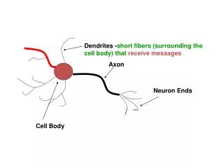
Axonal growth cone Karl H. Pfenninger Movement of the growth cone is mediated by a cytoskeletal lattice containing the motor proteins actin and myosin. As the neurite extends behind the moving growth cone, the microtubule backbone of the neurite is constructed from molecules of tubulin.
Guidance of Axons by short- and long-range cues: Attractive or repulsive Four types of mechanisms contribute to the guidance of the growth cone: Contact attraction, chemoattraction contact repulsion, chemorepulsion. Individual growth cones might be "pushed" from behind by a chemorepellent, "pulled" from in front by a chemoattractant, and "hemmed in" by attractive and repulsive local cues (cell surface or extracellular matrix molecules). Adapted from Tessier-Lavigne and Goodman (1996).
Molecular guidance moleculesConserved families of guidance molecules (A) and their receptors (B). Examples: ●SLIT secreted proteins, control midline repulsion, dual role, signaling through roundabout receptors (Robo) ● Ephrins (A +B) membrane anchored, repellent and attractive functions, receptors: EphA, EphB ●Netrins and their receptors ●Semaphorins 5 different subfamilies characterized by a 500 aa semaphorin domain, secreted and anchored. Cell Adhesion Molecules ( N-CAM, L1 or Fasciclins
Semaphorins example for dual function: Dual function: D) In the presence of NGF Sema III has a repellent effect on neurite growth E) In the presence of NT3, Sema III elicits outgrowth of neurites (NT-3) . Secreted (subclass 2 + 3) or membrane bound ligands (GPI anchored or transmembrane domain) have Chemorepellent or chemoattractive functions.
Linkage of the actin cytoskeleton to a permissive surface is required for forward advance. Actin is polymerized at the leading edge of the growth cone (right) and is swept toward the rear. If the actin meshwork is not linked to cell surface receptors that bind permissive molecules on adjacent cell surfaces, the actin cycles from front to rear but does not advance the growth cone. If the actin meshwork is attached to these receptors, the meshwork remains in place and newly polymerized actin helps advance the leading edge. Modified from Lin et al.(1994).
Ephrins +Eph receptors Ephrin-expressing cell (top) interacting with Eph-receptor expressing cell (bottom). Ligand–receptor interactions (green) are dimeric or oligomeric Eph–ephrin complexes. GPI, glycosylphosphatidylinositol; SAM, sterile alpha -motif. Functions: Vascular development Border formation Cell migration Axon guidance Synaptic plasticity Klas Kullander1 & Rüdiger Klein, 2002
Projections from preplate guide thalamocortical fibers Top: Preplate cells send out their axons towards the internal capsule (red). Thalamic axons project through the IC and meet cortical axons. Right: Handshake between thalamic and preplate axons and precise topography of early thalamicortical projections Note: Axons travel together (fasciculation) Axons use preexisting projections Guidepost cells show the way
Growth cones are sensory-motile organelles at the tip of growing axons and dendrites. Golgi-stained section of the spinal cord (specimen prepared by Ramon y Cajal, 1892, photographed 100 years later)
The cytoskeleton of the growth cone continuously changes during outgrowth and navigation.
Actin Tubulin
Growth cones are highly dynamic structures. Mauthner cell axon labeled with DiI in the spinal cord of a zebrafish embryo contacting a motoneuron (left) and forming an en passant synapse (right) Jontes et al., 2000
How does the growth cone get from A to B? Consider • Enormous distances. • Neuronal diversity.
Growth cones turn in response to gradients of axon guidance molecules Dickson, 2002
Four families of axon guidance molecules and their receptors • Netrins (DCC, Unc5) • Slits (Robo) • Semaphorins (plexin, neuropilin) • Ephrins/Eph (Eph/ephrin)
Gradient reading • requires detection of small concentration changes (a few percent over the length of the growth cone)
Gradient reading can be achieved by two mechanisms Netrin gradient n n n n n n n n n n n n n n n n n 1) Local autocatalysis (plus lateral inhibition) n n n n n 2) Adaptation Shallow, unreadable gradient Intracellularly enhanced gradient No gradient
Growth cone "sensory physiology" 1) Local autocatalysis amplifies a small concentration difference to generate a larger absolute difference. Lateral inhibition prevents the autocatalysis to spread and suppresses competing activation foci. 2) Adaptation shifts the baseline down to generate a larger relative concentration difference.
Growth cones are sensitive to external concentration differences of ca. 2% 1) Local autocatalysis (plus lateral inhibition) 300:100 102:100 3 : 1 2) Adaptation Shallow gradient Enhanced gradient
Calcium imaging with indicator dyes Fluo-3 Excitation wavelength (confocal Argon laser)
Growth cone guidance by local calcium increase and decrease Hi [Ca++]e Lo [Ca++]e Zheng, 2000
Caged calcium: Released by UV spot illumination of a growth cone loaded with NP-EGTA
Three examples of axon guidance in vivo 1) Navigation of commissural axons towards and across the midline. 2) Retinotectal map formation. 3) Olfactory system (if time allows).
Guidance across the midline • Conservation of mechanisms Dickson, 2002
Crossing the midline: Molecules and mechanisms Stein & Tessier-Lavigne, 2001
Crossing the midline:A smooth journey controlled by dynamic receptor interactions Stein & Tessier-Lavigne, 2001
Local protein synthesis is required for axon guidance beyond the midline Brittis et al., 2002
Morphogens BMP and Hedgehog in commissural axon guidance Charron et al., 2003
Genetic analysis of the retinotectal projection Optic nerve DiI injection DiO injection Chiasm Retina Optic tract Tectum
A screen for mutations disrupting axon pathfinding and retinotopyin zebrafish
Lipophilic axon tracers DiI (di-C18-...indo-carbocyanine) DiO (di-C18-oxa-carbocyanine) DiD (di-C18...-indo-di-carbocyanine) DiA (di-C16-…amino…styryl…pyrimidinium)
Axon pathfinding phenotypes discovered in the retinotectal screen Baier et al., 1996; Trowe et al., 1996; Karlstrom et al., 1996
The retinotectal projection creates a faithful map of the visual space in the brain D V (L) V D (M) N P (C) T A (R)
Sperry's chemoaffinity theory Connections between retinal and tectal neurons are specified by "key-and-lock" interactions of cell-surface molecules specific to these cells.
Positional information is graded and is being "read" by retinal axons 1. Growth cone guidance 2. Axon branching (not in all systems) 3. Refinement of axonal arbors
Attraction=Repulsion Axon guidance by gradients of attractive and repulsive cues in a two-dimensional field Branching D V Normal route Guidance from ectopic position A P
In vitro retinotectal guidance: The stripe assay Stripe assay was first carried out with crude membrane preparations from different parts of the tectum. ant post Walter et al., 1987
Stripe assay... ...was used to test molecules that were differentially expressed between anterior and posterior tectum. In 1995, the Bonhoeffer and Flanagan labs independently discovered the ephrins (under different names). Ephrin-A2 and ephrin-A5 are expressed as gradients in the tectum. Their receptors are expressed as gradients in the retina. ant: low ephrin-A post: high ephrin-A
Stripe assay results Ephrin-A2/mock Ephrin-A5 (1:2)/mock Ephrin-A5 (1:4)/mock Monschau et al., 1997
Ephrin-A2 and A5 both specify A/P position in the tectum assuming crowding results in a countergradient and/or more competition in anterior tectum Feldheim et al., 2000