Download
1 / 30
300 likes | 663 Views
Thromboembolic disease. -remains the third leading cause of direct maternal death -The pregnant woman increased risk of developing venous thromboembolism (VTE) compared with the non-pregnant which rises in the puerperium. Thromboprophylaxis in pregnancy.

E N D
Thromboembolic disease • -remains the third leading cause of direct maternal death • -The pregnant woman increased risk of developing venous thromboembolism (VTE) compared with the non-pregnantwhich rises in the puerperium
Thromboprophylaxis in pregnancy • Risk f a ct o r s f o r v e no us t hr o m bo e m bo lism • Pre-existing factors • Previous VTE • Family history of VTE (e.g. deficiency of protein C or S, antithrombin deficiency, prothrombin gene variant, Factor V Leiden) • Known thrombophilia • Lupus anticoagulant • Medical co-morbidities (e.g. sickle cell disease, cardiac disease, proteinuria >3 g/day) • Age >35 years • Obesity (BMI >30 kg/m2) • Parity >3 • Smoking • Intravenous drug user • Varicose veins
Obstetric factors • Pre-eclampsia • Dehydration/hyperemesis gravidarum • Multiple pregnancy • Caesarean section or forceps delivery • Prolonged labour • Postpartum haemorrhage • Transient factors • Systemic infection • Paraplegia or immobility • Recent surgical procedure • Ovarian hyper stimulation syndrome • Travel >4 hours • Midwife assessment on the following occasions: • initial meeting with the woman (booking visit) • any hospital admission • following the birth of the baby.
-If this assessment identifies women at risk of developing VTE, the midwife should promptly refer her to an expert in thrombosis in pregnancy • - commenced on subcutaneous injections of low molecular weight heparin (LMWH), as this does not cross the placental barrier with consequential effects on the fetus.
-midwife educate the woman in self-administration of the heparin, alerting her to carry a medical alert card containing such details with her at all times. • -provided with a sharps bin for safe disposal of the injection devices. • -Gradient compression stockings or TED stockings are likely to be prescribed,are available in two lengths, below the knee or thigh, and are designed to give a pressure gradient from the ankle to the knee or thigh
-Midwives should be trained in their use to be able to instruct the woman how to wear them correctly and monitor their use • -For hygiene purposes, stockings should be removed daily, but this should be no more than 30 minutes. • -The legs should be inspected and measured by the midwife every three days to detect any changes in size or tissue damage
-advice about avoiding dehydration, ceasing smoking and eating a healthy diet . • pregnant woman is expecting to travel long distances, especially by air, she will benefit by wearing loose fifing clothing and flight socks (TED stockings), drinking plenty of water, avoiding alcohol and remaining ambulant for as long as possible/performing leg exercises when at rest . • -During labour, encourage mobility with regular changes of position and passive leg exercises when the woman is at rest.
hydration is maintained and frequent examination of the woman's legs. • - If the woman has been prescribed LMWH in pregnancy, this should be omitted at the onset of contractions and regional anesthesia avoided within 12 hours of the last administered dose. • -There should be active management of the third stage of labour with the oxytoxic drug being administered IV.
-perineal suturing is required, avoid the woman being in the lithotomy position for a prolonged time, as this further increases the risk for deep vein thrombosis (DVT). • - If surgery is necessary, intermittent calf compression will be required in theatre. • -The postnatal period presents further risk to the woman for both DVT and pulmonary embolism (PE), and early mobilization should be encouraged. • postnatal observations, respiration rate and the development of any leg swelling. • - If either condition is suspected the woman must be referred urgently to a haematologist, or if at home she must be re-admitted to hospital.
Deep vein thrombosis • -A blood clot formed within a blood vessel is termed a thrombus, which can become detached and lodge in another blood vessel and partially or wholly occlude it. • -Virchow's triad -In pregnancy, Virchow's triad is affected by the physiological changes to the hematological system • -Despite pregnancy presenting a state of hypervolaemia, by term hypercoagulability also develops to compensate for the demands of the forthcoming labour and maintenance of haemostasis.
- venous stasis reaching its peak at 36 weeks and declining to pre-pregnancy values by 6 weeks following the baby's birth • the physical effect of the gravid uterus exerts pressure on the pelvic veins and the inferior vena cava, increasing the woman's risk of developing a DVT in the veins of the calf, thigh and pelvis
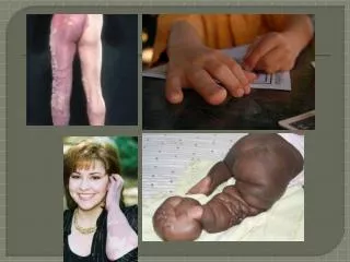
-In pregnancy, 90% of DVT occur in the left leg compared with 55% in the non-pregnant woman due to compression of the left iliac vein by the lt iliac artery in pregnancy • -The complications of DVT are pulmonary embolism (PE) and post-thrombotic syndrome
arising from damage to the venous valves that result in a backflow of blood, venous hypertension, oedema and tissue hypoxia. • -The midwife needs to be aware of the signs of DVT
pain in the area of the clot • swelling (usually one-sided) • red discoloration • difficulty in weight-bearing on the affected leg • low grade pyrexia • lower abdominal or back pain. • -If the leg appears swollen a tape measure should be used to assess the circumference of both legs at the affected area for comparison. • - A DVT is potentially life threatening and the midwife must refer the woman immediately to hospital for medical examination, investigation and treatment.
The classic diagnostic use of dorsiflexion of the foot (Homan's sign) is considered unreliable in pregnancy and the presence of severe lower back pain has greater significance • Doppler ultrasound and serum investigations might be performed • Venography is generally avoided in pregnancy due to the small radiation risk to the fetus. • -Treatment of DVT in pregnancy is with LMWH administered 12-hourly by subcutaneous injection to sustain the levels, and which should continue for at least 6 months after the diagnosis. • -Gradient compression stockings should be prescribed
-The woman will need to wear one on the affected leg for two years to reduce the risk of post-thrombotic syndrome • Anticoagulation therapy should continue for at least 6 months aher the diagnosis • -The woman should be seen by the anaesthetist prior to labour to discuss the risks that thromboembolic disorders have on the administration of regional/general anaesthesia. • - As soon as labour commences, heparin should be omitted and compression stockings should be worn. • - As regional anaesthesia carries a risk of spinal bleeding, this should be avoided within 12 hours of administration of heparin. • -Although general anaesthetic is itself a thrombotic risk, it may have to be considered for caesarean section.
encouraged to remain mobile • passive leg exercises • maintain hydration. • drugs given IV instead of intramuscularly (IM). • Prolonged use of the lithotomy position should be avoided, as this is a DVT risk. • - The third stage of labour should be actively managed with the oxytoxic drug being administered IV to prompt haemostasis. • - If perineal suturing is required, it should be undertaken promptly to limit the length of time the woman is in the lithotomy position -potential for PE during the postnatal period.
- woman who has had a previous DVT condition, encouraging early ambulation and hydration. • - Heparin is recommenced ,2 hours after a vaginal birth or longer if the woman had an epidural and/or caesarean section, and should continue until at least the 6-week postnatal appointment, at which point a decision to change to warfarin • -Oestrogen-based contraceptive pills are contraindicated so depo-provera or barrier methods of contraception should be discussed with the woman and her partner.
Pulmonary embolism • occurs when a DVT detaches and becomes mobile, known as an embolus. • - A large embolus might lodge in the pulmonary artery and smaller ones can travel distally to small vessels in the lung periphery, where they may wholly or partially occlude the blood vessel • impaired gaseous exchange • -After some hours, surfactant production by the affected lung ceases, the alveoli collapse and hypoxaemia results.
- Pulmonary arterial pressure rises and there is a reduction in cardiac output. • -The area of the lung affected by the embolism may become infracted
-In the case of a small embolism, there is likely to be: dyspnoea discomfort pain in the chest haemoptysis low grade pyrexia, all of which can be misdiagnosed as a chest infection. Cardiovascular examination is usually normal -A larger embolism that occludes a major vessel will result in a more acute presentation, because of sudden obstruction of the right ventricle and its outflow . There is : severe central chest pain due to ischaemiaPallor sweating as shock develops. Tachycardia occurs and a gallop rhythm of the heart may be heard on examination.
Hypotension develops as peripheral shutdown occurs. • Syncope may result when cardiac output is suddenly reduced • -Admission to an intensive care unit is highly likely as there is a significant risk of death if treatment is delayed. • -Pulmonary embolism is a medical emergency and urgent referral to hospital is indicated.
Diagnosis :clinical probability score and radiological imaging. • Heparin, usually LWMH, is commenced at presentation with subsequent anticoagulation treatment and management in labour and the postnatal period being similar to that for a woman presenting with DVT
Disseminated intravascular coagulation (DIC) • - DICdamage to the endothelium (lining of blood vessel walls) arising from pre-eclampsia, placental abruption, major haemorrhage, embolism, intrauterine fetal death or retained placenta results in thromboplastins being released from the damaged cells, causing the extrinsic pathway to mount a coagulation cascade. • -Blood clotting occurs at the original site and then small clots (micro-thrombi) formed • -Large quantities of fibrinogen, thrombocytes (platelets) and clotting factors V and VIII are consumed.
The micro-thrombi produced can occlude small blood vessels, resulting in ischaemia, hence some organ tissue dies and releases more thromboplastins and the cycle re-commences. • - All clotting factors and platelets are subsequently consumed and bleeding results. There is widespread blood clotting and a clotting deficiency. Bleeding occurs, petechiae develop in the skin and, if untreated, major haemorrhage can result