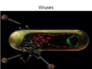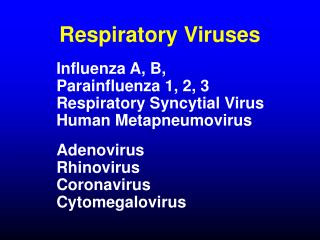

RESPIRATORY VIRUSES
E N D
Presentation Transcript
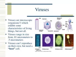
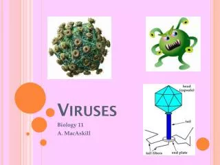
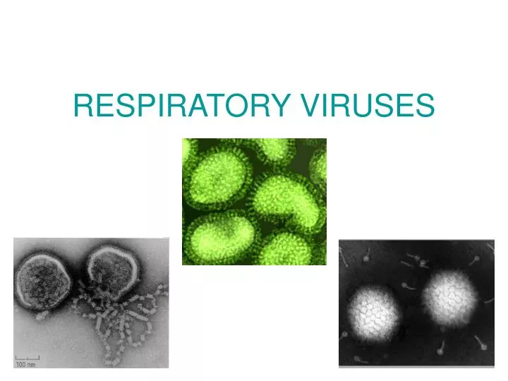
Family Orthomyxoviridae. Influenza virus • (-) RNA enveloped virus with haemagglutinin and neuraminidase spikes • Pleomorphic • Helical symmetry • Diameter 80-120 nm • 3 types: A, B and C
Structure of influenza virus • The internal antigens (matrix and RNP) are the type-specific proteins and are used to determine whether a particular virus is A, B, or C. • The external antigens (hemagglutininand neuraminidase) are subtype and strain- specific antigens of influenza A virus: H1N1, H2N2, H3N2
Hemagglutinin (HA) protein is involved in attachment and membrane fusion in the endosome of the infected cell.
The neuraminidase protein • digests sialic acid (neuraminic acid) on the surface of cells, when the virus binds to the cell, and it is internalized (endocytosed). • Promotes the release of virions from infected cells.
Antigenic variations • Antigenic drift is due to mutation. Both HA and NA after a few years may accumulate changes in genome that an individual immune to the original strain is not immune to the drifted one. Antigenic drift results in sporadic outbreaks and limited epidemics. • Antigenic shift is due to reassortment. It is the process by which at least two different strains of a virus combine to form a new subtype having a mixture of the surface antigens of the two original strains. As a result, the population has no immunity against the new strain. Antigenic shift results in an epidemic or pandemic. Antigenic drift creates influenza viruses with slightly modified antigens, while antigenic shift generates viruses with entirely novel antigens.
Past antigenic shift 1918H1N1 “Spanish Influenza” 20-40 million deaths 1957H2N2 “Asian Flu” 1-2 million deaths 1968H3N2 “Hong Kong Flu” 700,000 deaths • H1N1 Re-emergence No pandemic 2009H1N1 “Swine Flu Mild Pandemic • At least 15 HA subtypes and 9 NA subtypes occur in nature. • Up until 1997, only viruses of H1, H2, and H3 are known to infect and cause disease in humans.
Theories behind antigenic shift • 1. Reassortment of the H and N genes between human and avian influenza viruses trough a third host. • 2. Recycling of pre-existing strains(this probably occurred in 1977 when H1N1 re-surfaced). • 3. Gradual adaptation of avian influenza viruses to human transmission. Coinfection with human and animal strains of virus can generate very different virus strains by genetic reassortment
Reassortment of H and N genes between human and avian influenza viruses Sometimes pigs can be infected with more than one virus type at a time, which can allow the genes from these viruses to mix. This can result in an influenza virus containing genes from a number of sources, called a “reassortant” virus.
Bird or avian flu (2006) • The outbreaks affecting some Asian countries was caused by influenza A/H5N1 virus. • It caused severe infection in humans. • While avian influenza spreads rapidly among birds, it does not infect humans easily, and there is no confirmed evidence of human-to-human transmission.
2009 Influenza A (H1N1) Virus • The 2009 Swine flu outbreak in humans was due to a new strain of influenza A virus subtype H1N1 that derived in part from human influenza, avian influenza, and two separate strains of swine influenza(American swine and Eurasian swine viruses). • Swine influenza viruses are most commonly of the H1N1 subtype, but other subtypes are also circulating in pigs (e.g., H1N2, H3N1, H3N2).
Epidemiology and pathogenesis Spread: via small particle aerosols. • The incubation period is about 18 to 96 hours. • Site of infection: the epithelial cells of the respiratory tract: trachea and bronchi
Pathogenesis • Infection of mucosal cells results in cellular destruction and desquamation of the superficial mucosa. • The resulting edema and mononuclear cell infiltration are accompanied by local symptoms: nonproductive cough, sore throat, and nasal discharge. • Systemic symptoms: fever, muscle aches, headache,and general prostration.
Pathogenesis and immunity Immunityto an influenza virus is type-specific and lasts for many years. Recurrent cases of influenza are caused primarily by antigenically different strains.
LABORATORY DIAGNOSIS OF INFLUENZA Specimen: the nose and the throat swab. 1. Detection of antigenby immunofluorescence (IF) and ELISA. 2.The virus isolation in cell culture or chicken eggs. Identification of influenza strain and type: hemagglutination inhibition (HAI) and hemadsorption inhibition (HadsI). 3.Serology: HAI, neutralization test (NtT), ELISA, CFT in pared sera.
Immunofluorescence (IF) to detect virus into host cells direct IF indirect IF
Hemadsorption (Hads) • Virus growth in cell cultures is detected by testing for hemadsorption: red cells are added to the culture and adhere to virus budding from infected cells. • If the culture tests positive, hemadsorption inhibition test with specific antisera is used to identify the virus. cell culture positive Hads
Chick embryo culture method • Fluid from the amniotic or allantoic cavity of chick embryos is tested for the presence of newly formed viruses by haemagglutination test; • the virus in positive fluids is then identified by haemagglutination inhibition test with specific antisera. Inoculation of chick embryo Removing allantoicfluid
Haemagglutination inhibition test (HAI) Influenza viruses bind to red blood cells using the haemagglutinin causing the formation of a lattice. HA: two-fold serial dilutions of a virus are prepared, mixed with red blood cells, and added to the wells of a plastic plate. The red blood cells that are attached to virus particles form a lattice that coats the well. The red blood cells that are not bound by virus sink to the bottom of a well and form a button. The basis of the HAI assay is that antibodies to influenza virus will prevent attachment of the virus to red blood cells. By adding specific antibodies to the virus it is possible to block this interaction and detect the virus. If antibodies to the virus are specific, hemagglutination will not be observed. negative HA positive HA negative HAI positive HAI
Interferon • The innate immune system forms the first line of defence against viruses - Interferons (IFNs), glycoproteins known as cytokines. • Interferons are produced by: • the cells of the immune system (leukocytes, fibroblasts, or lymphocytes) in response to challenges by foreign agents such as viruses, parasites and tumor cells; • cells infected with a virus. • Interferons assist the immune response by: • inhibiting viral replication within host cells, • activating natural killer cells and macrophages, • inducing the resistance of host cells to viral infection.
VACCINES • The vaccine is multivalent and the current one is to two strains of influenza A and one of influenza B. • Inactivated preparation of egg-grown virus. • Live, attenuated vaccine. • Split-vaccine consists of all viral antigenes. • Sub-united vaccine consists of hemagglutinines and neuraminidases (H1N1, H3N2 and B).
Unique features of Adenoviruses • Double-stranded linear(+)DNA. • Naked icosahedral capsid has fibers (viral attachment proteins) and vertices. • 47 human serotypes • Viruses cause: • lytic, • persistent, • latent infections in humans, • some strain can immortalize certain animal cells.
Pathogenesis of adenoviruses infections • Virus is spread: • by aerosol, • direct contact, • fecal-oral. • Virus infects: epithelial cells in respiratory and gastrointestinal tract, conjunctiva and cornea. • Virus persists in lymphoid tissue (tonsils, adenoids, Peyer’s patches).
ILLNESS ASSOCIATED WITH ADENOVIRUSES • Incubation period is 2-14 days.Clinical syndromes: • Eye Epidemic keratoconjunctivitis, acute follicular conjunctivitis, pharyngoconjunctival fever. • Respiratory system Common cold (rhinitis), pharyngitis, tonsillitis, bronchitis, pneumonia. • Genitourinary Acute hemorrhagic cystitis, orchitis, nephritis. • Gastrointestinal Gastroenteritis, mesenteric adenitis, appendicitis. • Rare results of adenovirus infections: Meningitis, encephalitis, arthritis, myocarditis, hepatitis. Fatal disease may occur in immunocompromised patients, as a result of a new infection or reactivation of latent virus
IMMUNITY Strong, type-specific PREVENTION A vaccine is available against Adult Respiratory Distress Syndrome. Oral, live-attenuated vaccines against serotypes 4 and 7 of adenovirusis administered in tablet form. It is given to new recruits into various arm forces around the world.
LABORATORY DIAGNOSIS OF ADENOVIRUSES INFECTION 1. Detection of antigenfrom pharyngeal and eye secretions and feces by: IF, ELISA and polymerase chain reaction. 2. The virus isolation in cell culture. Cytopathic effects (CPE) include swelling and rounding of cells. Identification: HAI, CFT; type virus – by NtT. 3. Serology (rise in antibody titer): HAI, NtT, CFT.
Family Paramyxoviridae • GenusRespirovirus(parainfluenza viruses types1 and 3) • Genus Rubulavirus (parainfluenza viruses types 2 and 4, mumps virus) • GenusMorbillivirus(measles virus) • GenusPneumovirus (respiratory syncytial virus)
Parainfluenza viruses • ss (-) RNA viruses • Enveloped viruses with hemagglutinin and neuraminidase spikes and fusion (F) protein • Helical symmetry • 4 types: 1, 2, 3, 4a, 4b
Clinical features of parainfluenza (PIV) • Incubation period is 2 to 6 days. • Clinical symptoms: • Rhinitis, pharyngitis, cough, fever, croup (laryngotracheobronchitis), bronchiolitis, and pneumonia. • Croup- the subglottic region becomes narrower and results in difficulty with breathing, a seal bark-like cough and hoarseness. • There is clinical variation between the different PIV types. • PIV-1 and 2: croup in children ages 2-6 years in autumn/early winter. • PIV-3: bronchiolitis and pneumonia, andcroup sporadically, without a particular seasonal occurrence. • PIV-4: mild upper respiratory infections.
LABORATORY DIAGNOSIS OF PARAINFLUENZA 1. Detection of antigen from nasopharyngeal aspirates and throat swab by IF and PCR. 2. The virus isolation in cell culture. Indication:Haemadsorption of erythrocytes on the surface of cells infected with virus. Identification: HadsI, HAI, NtT, CFT. 3. Serology – detection of rise in titer of IgGin pared sera: NtT, ELISA, CFT, HAI.
Respiratory Syncytial Virus (RSV) • ss RNA enveloped virus • Considerable strain variation exists, may be classified into subgroup A and B. • CPE - the formation of multi-nucleate syncytium Clinical features:
MUMPS • Mumps is a viral infection that primarily affects the salivary glands causing them to become inflamed, resulting in the characteristic “chipmuck cheeks” • Transmission by: • salivary gland secretions, mainly just before and shortly after clinical onset • direct and close person-to-person contact and • airborne route. The virus enters the body through the pharynx or the conjunctiva.
Clinical symptoms of mumps • The incubation period usually is 18 to 21 days, but may extend from 12 to 35 days. • Prodromal phase:fever, anorexia, malaise, myalgia. • The symptoms: • Fever. • Parotitis. Pain from parotitis swelling persists for 7 - 10 days. It may be unilateral or bilateral. • Up to 20% infections result in no symptoms.
Complications of mumps • Orchitis 20-50 % • Meningitis and meningoencephalitis 15 % • Ovaritis 5 % • Pancreatitis 2-5 % • Rare complications: polyarthritis, diabetes, nephritis, thyroiditis, deafness, myocarditis.