Download
1 / 41
430 likes | 736 Views
CARBOHYDRATE METABOLISM. Carbohydrate metabolism includes. Digestion of carbohydrates. Absorption of digested carbohydrates. Utilization of carbohydrates. Utilization. A-Anabolic pathways :

E N D
Carbohydrate metabolism includes Digestion of carbohydrates. Absorption of digested carbohydrates. Utilization of carbohydrates
Utilization A-Anabolic pathways: Transforming small molecules into big molecules constituting the body structures and machinery. It is energy requiring, e.g., glycogenesis and lactose synthesis B-Catabolic pathways: Breakdown of large molecules into smaller molecules to produce energy or smaller molecules or reducing equivalents, e.g., HMP-shunt and uronic acid pathway. C-Amphibolic pathways: They are utilized for both anabolic and catabolic purposes, e.g., glycolysis and Krebs' cycle.
Digestible carbohydrates: starch, glycogen, maltose, sucrose, and lactose (oligosaccharides and polysaccharides). Ready-to-absorb carbohydrates: Which do not need digestion and are absorbed as such, e.g., monosaccharides: glucose, mannose, galactose, fructose and pentoses. Non-digestible carbohydrates: Which are called dietary fibers. These include cellulose, gums and pectins. They are very important for stools excretion and are anticancer and intestinal bacteria feed on it to release certain vitamins.
Carbohydrate Digestion in the buccal cavity: In the mouth, salivary amylase is produced by the salivary glands. Its optimum pH is 6.7 - 6.8 and is activated by chloride ions. It is a -glycosidase specific for hydrolysis of -1,4 glucosidic linkage present in cooked starch and glycogen producing maltose and dextrins. Salivary amylase cannot digest -1,4-glucosidic linkage in cellulose.
2. Carbohydrate digestion in the Stomach: Salivary amylase continues to act on starch, glycogen or dextrins for 2 - 3 minutes only in the stomach (acidic pH 1 - 2).
3. Carbohydrate digestion in the Small Intestine: In the small intestine, there are two juices that digest carbohydrates: A. The pancreatic juice: This juice contains pancreatic amylase, an -glycosidase. It has an optimum pH 7.1 and is also activated by chloride ion. It acts exactly as salivary amylase, digesting cooked and uncooked starch, glycogen and starch dextrins which escaped digestion by salivary amylase in the mouth producing maltose, maltotriose (three -glucose residues linked by -1,4 bonds) and a mixture of branched oligosaccharides (-limited dextrins), non-branched oligosaccharides and some glucose.
B. Intestinal mucosal brush border enzymes: The final digestion of carbohydrates occurs in the small intestine by the action of the following disaccharidases: Lactase hydrolyzes lactose into glucose and galactose. Lactase deficiency leads to lactose intolerance, an inherited or age-dependent decline of enzyme expression or an acquired medical problem due to intestinal diseases such as colitis and gastroenteritis. Symptoms include abdominal cramps, diarrhea, and flatulence on eating fresh and non-fermented milk products. It is treated by consumption of live-culture yogurt.
Dietary fibers The -1,4 glucosidic linkage of Cellulose is not hydrolyzed by human digestive enzymes. Hemicellulose, gums, pectins and pentosans are also indigestible. Cellulose and other dietary fibers passes as it is in stools, increasing bulk of intestinal contents by adsorbing water and stimulates peristaltic movements to reduces stool transit time and prevents constipation. They bind and dilute bile acids. The more soluble fibers found in legumes and fruit, e.g., gums and pectins, lower blood cholesterol, possibly by binding bile acids and dietary cholesterol.
The soluble fibers also slow the stomach emptying and attenuate the post-prandial rise in blood glucose that spares insulin. This effect is beneficial to diabetics and to dieters because it reduces the rebound fall in blood glucose that stimulates appetite It induces establishment of normal colon bacteria with several benefits including fermentation of fibers and production of vitamins (e.g., vit. K)
Carbohydrates Absorption
Site of absorption: Mainly the upper part of small intestine, i.e., jejunum. Very small amount is absorbed in the stomach or large intestine.
Route of absorption By the portal vein to the liver, i.e., blood stream chiefly in the form of hexoses (glucose, fructose, mannose and galactose) and as pentose sugars (ribose).
Mechanism of absorption (or transport): A) Facilitated absorption: This mechanism is the most active for fructose and pentoses but is utilizable by glucose and galactose if their lumenal concentration is favorable. Absorption is derived by concentration gradient of sugar in the intestinal lumen, i.e., sugars passes from high concentration in lumen to lower concentration in mucosal cells then to blood. It utilizes a sodium-dependent facilitated glucose transporter, Glut5 and sodium-independent for fructose and pentoses.
B) Active absorption: This absorption occurs against concentration gradient, i.e., sugars are absorbed from low to high concentration and requires the presence of an OH group on C2 at the right side of a pyranose ring and a methyl group or a substituted methyl group at C5. This applies to glucose and galactose. It utilizes a sodium-dependent glucose transporter, SLGT1. A mobile carrier protein molecule (glucose transporters) presents in the cell membrane of all cells including intestinal cells.
Glucose transporter has 2 sites, one for sodium and the other for glucose, symporting sodium down its concentration gradient and glucose against its concentration gradient across cell membrane. Both sodium and glucose are released within mucosal cells, allowing the carrier to recycle for more cargo. Sodium is pumped out again by ATP-dependent Na+-K+-exchange pump. The ratio of Na+/glucose transported varies according to type of transporter to be 1:1 or 3:1 ratio. There is also an Na+-independent glucose transporter, Glut2, that transports glucose and other hexoses out of mucosal and other cells into blood
http://www.youtube.com/watch?v=6xqf6-RH6nk http://www.youtube.com/watch?v=yz7EHJFDEJs
Fate of glucose A-Oxidative fate: Major pathways: A. Glycolysis. B. Krebs' cycle. Minor pathways: A. Pentose shunt. B. Uronic acid pathway. B- Anabolic fates: Glycogenesis/glycogenolysis. Gluconeogenesis. Monosaccharides synthesis. Lactose synthesis. Glycolipids, glycoproteins and Proteoglycans synthesis.
Major Oxidative Pathways of Glucose: Glycolysis Embden-Myerhof Pathway Anaerobic Oxidation of Glucose
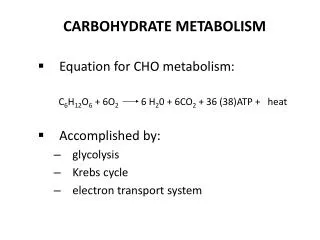
Definition: It is a cascade of reactions that converts glucose into two pyruvate molecules or into lactate aiming at production of ATP and other intermediates. It is also utilized in its opposite direction in gluconeogenesis.
Intracellular site and tissue distribution: It occurs in the cell cytosol of all tissues of the body. 1.RBCs: are devoid of mitochondria and depend on glycolysis as the main source of energy. Mammalian erythrocyte is unique in that about 90% of its total energy requirement is provided by glycolysis. 2.Contracting muscles due to occlusion of blood vessels by the muscular contraction that decreases oxygen 3-Cornea, lens and some parts of retina which have a limited blood supply and lack mitochondria which if present would absorb and scatter light interfering with transparency.
4-Kidney (medulla), testicles, leukocytes and white muscle fibers, where there are relatively few mitochondria. 5-Cancer cells due to dissociation of the high rate of glycolysis from Krebs', i.e., aerobic production of lactate. 6-Brain and gastrointestinal tract also normally derive most of their energy from glycolysis.
Biological importance (or Functions) of glycolysis: Glucose oxidation producing ATP. It is the major source of energy in certain tissues, e.g., RBCs and skeletal muscles. It provides pyruvic acid needed for Krebs' cycle. It is a link with fat metabolism, e.g., dihydroxyacetone phosphate into glycerol 3-phosphate in adipose tissue. It a link with amino acid metabolism, e.g., 3-phopshoglycerate into serine and pyruvate into alanine and vice versa. Production of 2,3-DPG that is important in tissue oxygenation.
It is the major source of lactic acid that is gluconeogenic. Reversal of glycolysis is gluconeogenesis, an important source of glucose. Main pathway of metabolism of fructose from the diet. A small number of genetic diseases occur due to deficiency in activity of enzymes ofglycolysis, are manifested mainly as hemolytic anemias. Cancer cells are glycolytic producing large amount of lactate, favoring a relatively acidic local pH in the tumor, a situation that was utilized to develop therapy for cancer that could be locally activated by this acidic pH.
It is oxidation of glucose (or glycogen in muscles) into pyruvate when oxygen is available for respiratory chain or lactate in the absence of oxygen. Glucose is transported into cells by glucose transporter (at least 6 types) that is insulin dependent in muscles and adipose tissue but not in other vital tissues, e.g., brain, heart, kidney and RBCs. Liver is freely permeable to glucose in and out but insulin activates glucose utilization in liver by activating glucokinase.
Under anaerobic conditions: 1- Total ATP lost = 2 ATP as follows, One ATP in the activation of glucose to glucose-6-phosphate. One ATP in the activation of fructose-6-phosphate to fructose1,6-diphosphate. 2- Total ATP gained = 4 ATP as follows, 2 ATP by substrate level phosphorylation from 1,3-diphosphoglycerate 2 ATP from substrate level phosphorylation from phosphoenol pyruvate. 3- Net ATP gained = 4 ATP gained - 2 ATP lost = 2 ATP for the anaerobic oxidation of one mole of glucose into lactate.
Under aerobic conditions: Total ATP lost = 2 ATP. Total ATP gained = 10 ATP are generated as follows, 4 ATP (obtained by substrate level phosphorylation) + 2 NADH.H+ chain (produced from oxidation of glyceraldehyde-3-phosphate) 2 X 3 ATP = 6 ATP, after oxidation in the functioning respiratory Net ATP gained = 8 ATP as follows, 10 ATP – 2 ATP = 8 ATP for the aerobic oxidation of one mole of glucose.
A. Key regulatory enzymes: are those enzymes that catalyze the irreversible steps of glycolysis that include three steps as follows, 1-Phosphofructokinase: It is an allosteric enzyme stimulated by high levels of fructose-6- phosphate, fructose-2,6-diphosphate (in liver), ADP and AMP, Pi, and ammonia. It is inhibited allosterically by ATP, low pH and citrate.
2-Hexokinase: Accumulation of glucose-6-phosphate and inhibition of phosphofructokinase results in accumulation of fructose-6-phosphate and glucose-6-phosphate that allosterically inhibit hexokinase. 3-Pyruvate kinase: It is inhibited also by excess ATP, fatty acids, and acetyl-CoA and is stimulated by fructose-1,6-diphosphate, ADP and AMP It is regulated by cAMP-dependent phosphorylation-dephosphorylation mechanism
B. Hormonal regulation: 1. Insulin: Stimulates synthesis of glucokinase, phosphofructokinase and pyruvate kinase, so it stimulates glycolysis. It also induces glucose transporters to provide cells with glucose for glycolysis. 2-Adrenaline and glucagon are inhibitory by inhibiting pyruvate kinase.
Thank You Edited by Dr/Ali H. El-Far Lecturer of Biochemistry Fac. of Vet. Med. Damanhour Univ.