Download
1 / 53
590 likes | 703 Views
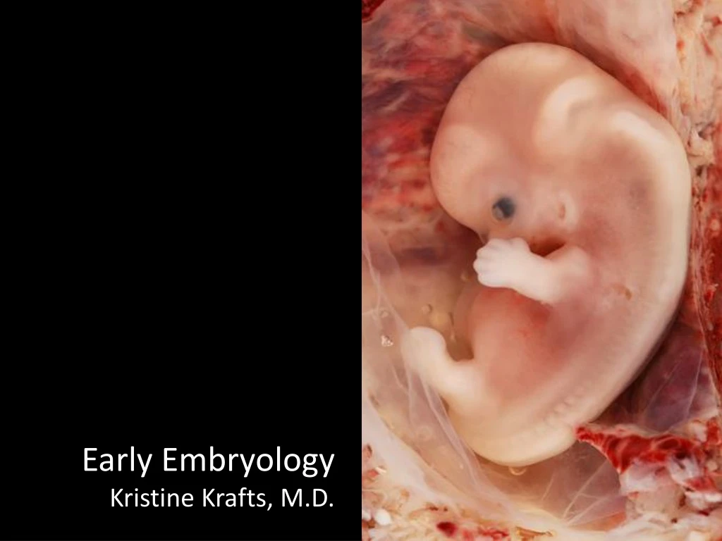
Explore the key stages of embryonic development in this informative lecture. Learn about the phases, gastrulation process, and neural tube formation. Dive into the fate of germ cell layers and pharyngeal arches, along with clinical correlations like Treacher Collins Syndrome. Discover the significance of folding in embryo development. For a visual guide, check out the YouTube video linked in the outline.

E N D
Embryology Lecture Objectives • In general, what happens in the first and second phases of the embryonic period? • What happens during week 1 and 2? • Describe the main events in gastrulation. • How does the neural tube form, and what happens to it?
Embryology Lecture Objectives • What is the fate of the endoderm, mesoderm and ectoderm? • What are the pharyngeal arches? What structures arise from each arch, groove, and cleft? • What structures do neural crest cells form? major stuff forms from pouches?
Embryology Lecture Outline • General overview of prenatal development • Embryonic period phase 1 • Formation of bilaminar disk • Formation of trilaminar disk (gastrulation) • Embryonic period phase 2 • Formation of neural tube • Differentiation of mesoderm • Folding of embryo • Formation of pharyngeal arches
Embryology Lecture Outline General overview of prenatal development
Prenatal Development Embryo Fetus Fertilization Phase 1 Cellular proliferation and migration Phase 2 Differentiation of internal & external structures Phase 3 Growth and maturation 0 1 2 3 4 5 6 7 8 40 Delivery
This YouTube video is awesome at explaining early embryonic development: http://www.youtube.com/watch?v=rN3lep6roRI
Embryology Lecture Outline General overview of prenatal development Embryonic period phase 1
Prenatal Development Embryo Fetus Fertilization Phase 1 Cellular proliferation and migration Phase 2 Differentiation of internal & external structures Phase 3 Growth and maturation 0 1 2 3 4 5 6 7 8 40 Delivery
Embryology Lecture Outline • General overview of prenatal development • Embryonic period phase 1 • Formation of bilaminar disk
Week 1: Differentiation of Morula into Blastocyst Morula Blastocyst
Week 2: Formation of Bilaminar Germ Disk epiblast hypoblast
Embryology Lecture Outline • General overview of prenatal development • Embryonic period phase 1 • Formation of bilaminar disk • Formation of trilaminar disk (gastrulation) "It is not birth, marriage, or death, but gastrulation which is truly the most important time in your life.” - Lewis Wolpert (1986)
Gastrulation: formation of primitive streak primitive streak primitive node epiblast
Gastrulation: movement and differentiation of epiblast cells Bilaminar germ disk Primitive streak Epiblast Hypoblast Endoderm Ectoderm Mesoderm Endoderm Epiblast cells give rise to all three germ cell layers!(the hypoblast does NOT turn into endoderm)
Gastrulation: formation of notochord The notochord is super important because it tells the three layers what to do next.
Embryology Lecture Outline • General overview of prenatal development • Embryonic period phase 1 • Formation of bilaminar disk • Formation of trilaminar disk (gastrulation) • Embryonic period phase 2
Prenatal Development Embryo Fetus Fertilization Phase 1 Cellular proliferation and migration Phase 2 Differentiation of internal & external structures Phase 3 Growth and maturation 0 1 2 3 4 5 6 7 8 40 Delivery
Embryology Lecture Outline • General overview of prenatal development • Embryonic period phase 1 • Formation of bilaminar disk • Formation of trilaminar disk (gastrulation) • Embryonic period phase 2 • Formation of neural tube
Embryology Lecture Outline • General overview of prenatal development • Embryonic period phase 1 • Formation of bilaminar disk • Formation of trilaminar disk (gastrulation) • Embryonic period phase 2 • Formation of neural tube • Differentiation of mesoderm
Differentiation of mesoderm Paraxial mesoderm forms bones and muscles of most of the body (except head), pharyngeal arches, and connective tissue Intermediate mesoderm forms urogenital system Lateral plate mesoderm forms hematopoietic system, heart, pharyngeal arches, and connective tissue
Central nervous system Cranial nervesBones and connective tissue of head Pharyngeal arches Neuroectoderm Urogenital system Neural crest Intermediate plate mesoderm Know this! Lateral plate mesoderm Surface ectoderm Paraxial mesoderm Epidermis Heart Hematopoietic system Pharyngeal arches Connective tissue Bones of most of the body (everything except the head)Muscles of the body and head Pharyngeal arches Connective tissue Endoderm Lining of GI tract
Clinical Correlation: Treacher Collins Syndrome Neural crest cells don’t migrate properly to the facial region. Structures derived from 1st and 2nd pharyngeal arches don’t develop properly.
Embryology Lecture Outline • General overview of prenatal development • Embryonic period phase 1 • Formation of bilaminar disk • Formation of trilaminar disk (gastrulation) • Embryonic period phase 2 • Formation of neural tube • Differentiation of mesoderm • Folding of embryo
Lateral Folding of the Embryo Lateral plate mesoderm splits in two. One part remains near the ectoderm. The other part follows the endoderm.
Lateral Folding of the Embryo Endodermal layer (lined by mesoderm) bends, the edges reaching towards each other, meeting in front to form the gut. Ectodermal layer (lined by mesoderm) grows forward, reaches around the gut, and zips up the front to form the anterior body wall.
Lateral Folding of the Embryo Back, with ectoderm overlying neural tube Gut (lined by endoderm, surrounded by mesoderm) Amnion (and amniotic cavity) comes along for the ride, eventually surrounding entire embryo. Anterior thoracic wall (mesoderm covered with ectoderm)
Head-Tail Folding of the Embryo Day 24 Before folding Stomatodeum Day 26 Day 28
What happens to the neural tube? It turns into the brain(forebrain, midbrain and hindbrain) and the spinal cord. • Hey, what’s that mesoderm doing there? It’s forming: • Somatomeres (which turn into muscles of the head and neck) • Somites (which turn into the bones and muscles of the back).
Embryo, day 23-26 Neural tube still open Neural tube closed Somites Neural tube still open
Embryo, day 26-30 Neural tube closed Somites Neural tube still a little bit open
Embryology Lecture Outline • General overview of prenatal development • Embryonic period phase 1 • Formation of bilaminar disk • Formation of trilaminar disk (gastrulation) • Embryonic period phase 2 • Formation of neural tube • Differentiation of mesoderm • Folding of embryo • Formation of pharyngeal arches
Pharyngeal Arch Anatomy Arches have a core that’s derived from lateral and paraxial mesoderm and neural crest cells. They are covered with ectoderm on the outside and lined by endoderm on the inside. Each arch has its own cartilage, artery, and nerve.
Formation of Pharyngeal Arches 25-day-old embryo 35-day-old embryo No arches yetBuccopharyngeal membrane intact Arches and pouches nicely formed Mouth now open to esophagus
Embryo, day 26-30 Frontal prominence 1 2 3 Somites
Optic placode Nasal placode Stomatodeum Maxillary 1 Mandibular Heart 2 3 4 Somites First four arches in a 32-day-old embryo
Wait, are they branchial or pharyngeal arches? Fish have branchial (“gill”) arches, with slits in between for water flow. Humans don’t have gills. So our arches are called pharyngeal.
Groove/cleft Arch Pouch Pharyngeal arches, grooves/clefts and pouches
Know the stuff in red! Important! Meckel’s cartilage indicates where the mandible will develop – but it does not turn into the mandible!
What happens to the pouches and grooves? • 1st groove and pouch -> ear stuff • Rest of grooves disappear • 2nd pouch obliterated by tonsil • 3rd pouch -> inferior parathyroid, thymus • 4th and 5th pouches -> superior parathyroid, ultimobranchial body
Head and Neck Anomalies From Improper Groove Closure Pharyngeal sinuses and cysts Auricular sinuses and cysts
Prenatal Development Embryo Fetus Fertilization Phase 1 Cellular proliferation and migration Phase 2 Differentiation of internal & external structures Phase 3 Growth and maturation 0 1 2 3 4 5 6 7 8 40 Delivery We covered this in this lecture.