Download
1 / 40
400 likes | 425 Views
Explore the anatomy and functions of the cerebellum, a crucial brain structure responsible for motor coordination and learning. Learn about its different lobes, neural arrangement, input pathways, and motor outputs.

E N D
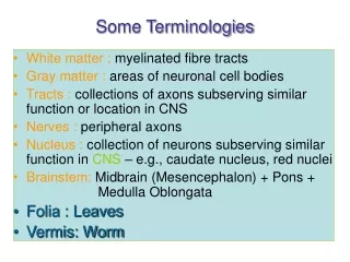
Some Terminologies • White matter : myelinated fibre tracts • Gray matter : areas of neuronal cell bodies • Tracts: collections of axons subserving similar function or location in CNS • Nerves: peripheral axons • Nucleus : collection of neurons subserving similar function in CNS – e.g., caudate nucleus, red nuclei • Brainstem:Midbrain (Mesencephalon) + Pons + Medulla Oblongata • Folia : Leaves • Vermis: Worm
Brain components Table 5.3 (1)Page 144 Cerebral cortex Cerebral cortex Basal nuclei (lateral to thalamus) Basal nuclei Thalamus (medial) Thalamus Diencephalon Hypothalamus Hypothalamus Cerebellum Cerebellum Midbrain(Mesencephalon) Brain stem (midbrain, pons, and medulla) Brain stem Pons Medullaoblongata Spinal cord
The Cerebellum • Located dorsal to the pons and medulla • Makes up 11% of the brain’s mass • Cerebellar activity occurs subconsciously • Provides precise timing and appropriate patterns of skeletal muscle contraction Programming ballistic movements • Acts as comparator for movementsComparing intended and actual movement • Correction of ongoing movementsInternal & external feedbackDeviations from intended movement • Motor learningShift from conscious ---> unconscious
Anatomy of the Cerebellum 2 symmetrical hemispheres connected medially by the Vermis Folia: Transversely oriented gyri 3 lobes in each hemisphere:Anterior, Posterior, Flocculonodular (FN) Neural arrangement:Gray matter (Cortex), White matter (Internal), Scattered cerebellar nuclei: dentate, globose, emboliform, fastigial Arbor vitae (tree of life): distinctive treelike pattern of the white matter Folium
Vestibulocerebellum Spinocerebellum Cerebrocerebelum Cerebellum Primary fissure Anterior Lobe Regulation of muscle tone, coordination of skilled voluntary movement Posterior Lobe Planning and initiation of voluntary activity Flocculo-Nodular Lobe (FN lobe) Maintenance of balance, control of eye movements Folia
Intermediate part Lateral part Cerebellum
Cerebellum: the Structure Inputs to the cerebellar cortex: Climbing fibers & Mossy fibers Climbing fibers: originate in the inferior olive of the medulla Mossy fibers: originate in all the cerebellar afferent tracts apart from inferior olive Purkinje cells: The final output of the cerebellar cortex 3 Layered CerebellarCortex
Cerebellum: 3 layered cortex Climbing fibers: excite the Purkinje cells Mossy fibers: excite the granule cells Granule cells: make excitatory contact with the Purkinje cells Purkinje cells: Tonic inhibition on the activity of the neurons of the cerebellar nuclei => All excitatory inputs will be converted to the inhibition => Removing the excitatory influence of the cerebellar inputs (erasing)
CerebellarPeduncles Cerebellar Peduncles • Three paired fiber tracts connect the cerebellum to the brainstem: • Superior peduncles connect the cerebellum to the midbrain; • Middle peduncles connect the cerebellum to the pons and to the axis of the brainstem; • Inferior peduncles connect the cerebellum to the medulla.
Cerebellar Peduncles Superior peduncles (to the midbrain): Fibers originate from neurons in the deep cerebellar nuclei & communicates with the motor cortex via the midbrain and the diencephalon (thalamus) Middle peduncles (to the pons): Cerebellum receives information advising it of voluntary motor activities initiated by motor cortex Inferior peduncles (to the medulla): Afferents conveying sensory information from muscle proprioceptors throughout the body & from the vestibular nuclei of the brainstem (Spinal cord)
2 maps of body Primary fissure Inputs to cerebellum from spinocerebellar tracts have a somatotopic organization. Signals from the motor cortex, which is also arranged somatotopically, project to corresponding points in the sensory maps of the cerebellum.
Cerebellar Inputs • Vermis • Receives input from spinal cord regarding somatosensory and kinesthetic information (intrinsic knowledge of the position of the limbs) • Damage leads to difficulty with postural adjustments (cerebellar ataxia) • Intermediate Zone • Receives input from the red nucleus and somatosensory information from the spinal cord • Damage results in rigidity & difficulty in moving limbs • Lateral Zone • Receives input from the motor and association cortices through the pons • Projects to the dentate nucleus, which projects back to primary and premotor cortex • Damage leads to 4 types of deficits:- Ballistic movements (cerebellar ataxia) - Coordination of multi-joint movement (lack of coordination: asynergia)- Muscle learning (loss of muscle tone: hypotonia)- Movement timing
Outputs of the Cerebellum Cerebellar nuclei: dentate, globose, emboliform, fastigial Dentate nuclei: project contralaterally through the superior cerebellar peduncle to neurons in the contralateralthalamus & from thalamus to motor cortexFunc.: influence planning and initiation of voluntary movement Emboliform & Globose nuclei: project mainly to the contralateral red nuclei & a small group is projected to the motor cortexRed Nuclei Rubrospinal Tract control of proximal limb muscles Fastigial nuclei: project to the vestibular nuclei & to the pontine and medullaryreticular formation Vestibulospinal & Reticulospinal tracts
Clinical Findings and Localization of Cerebellar Lesions Ataxia refers to disordered contractions of agonist and antagonist muscles and lack of coordination between movements at different joints typically seen in patients with cerebellar lesions. Normal movements require coordination of agonist and antagonist muscles at different joints in order for movement to have smooth trajectory. In ataxia movements have irregular, wavering course consisting of continuous overshooting, overcorrecting and then overshooting again around the intended trajectory. Dysmetria = abnormal undershoot or overshoot during movements toward a target (finger-nose-finger test).
Cerebellum and Motor Learning • Deficits in learning complex motor tasks after cerebellar lesions • fMRI studies : cerebellum active during learning of novel movements • Postulated that cerebellar nuclei store certain motor memories • May be involved in cognitive functions
Cerebellum: Control of Voluntary Movement Cerebellum has no direct connection to the spinal motoneurons (indirect effect). • Information sources: lesions & damages & experimental stimulation of cerebellar nuclei • Primary function: • To supplement & correlate the activities of other motor areas • Control of posture • Correction of rapid movements initiated by cerebral cortex • Motor learning Frequency of nerve impulses in the climbing fibers almost doubles when a monkey learns a new task • Movement Control: • Inputs from motor cortex inform the cerebellum of an intended movement before it is initiated • Sensory information is then received via the spinocerebellar tract • An error signal is generated and is fed back to the cortex
Cerebellar Processing • Cerebellum receives impulses of the intent to initiate voluntary muscle contraction • Proprioceptors and visual signals “inform” the cerebellum of the body’s condition • Cerebellar cortex calculates the best way to perform a movement • A “blueprint” of coordinated movement is sent to the cerebral motor cortex Cerebellar Cognitive Function • Plays a role in language and problem solving • Recognizes and predicts sequences of events
Table 5.3 (1)Page 144 Thalamus (medial) Hypothalamus Diencephalon • Central core of the forebrain • Consists of three paired structures – thalamus, hypothalamus, and epithalamus • Encloses the third ventricle
Table 5.3 (1)Page 144 Thalamus (medial) Hypothalamus Thalamus • Paired, egg-shaped masses that form the superolateral walls of the third ventricle • Contains four groups of nuclei : anterior, ventral, dorsal, and posterior • Nuclei project and receive fibers from the cerebral cortex
Thalamic Function • Afferent impulses from all senses converge and synapse in the thalamus • Impulses of similar function are sorted out, edited, and relayed as a group • All inputs ascending to the cerebral cortex pass through the thalamus • Plays a key role in mediating sensation, motor activities, cortical arousal, learning, and memory
Table 5.3 (1)Page 144 Thalamus (medial) Hypothalamus Hypothalamic Nuclei
Hypothalamic Function • Regulates blood pressure, rate and force of heartbeat, digestive tract motility, rate and depth of breathing, and many other visceral activities • Is involved with perception of pleasure, fear, and rage • Controls mechanisms needed to maintain normal body temperature • Regulates feelings of hunger and satiety • Regulates sleep and the sleep cycle • Endocrine Functions of the Hypothalamus
Central sulcus Frontal lobe Parietal lobe Parietooccipital notch Occipital lobe Lateral fissure Preoccipital notch Temporal lobe The Cerebral Cortex
The cerebral cortex • Cerebral Cortex : outer layer of gray matter • It covers an inner core of white matter • The gross structure has gyri and sulci Different Lobes: • Frontal : voluntary motor activity, speaking ability, and elaboration of thought; stimulation of different areas of its primary motor cortex moves different body regions, again primarily on the opposite side of the body. • Parietal : somatosensory processing; each region of its cortex receives somaesthetic and proprioceptive input from a specific body area, primarily from the opposite body side. • Temporal : receives sound sensation • Occipital : initial processing of visual input
Somatosensory cortex (Somesthetic sensation and proprioception) Supplementary motor area (programming of complex movement) Primary motor cortex (Voluntary movement) Premotor cortex (coordination of complex movements) Posterior parietal cortex (integration of somatosensory and visual input) Central sulcus Prefrontal association cortex (planning for voluntary activity; decision making; personality traits) Parietal lobe Wernicke’s area (speech understanding) Frontal lobe Parietal-temporal-occipital association cortex (integraton of all sensory input- imp in language) Broca’s area (speech formation) Primary auditory cortex Occipital lobe Limbic association cortex (motivation, emotion, memory) Temporal lobe Primary visual cortex
Parietal Lobe - Somatosensory cortex • Somesthetic sensation - sensations from the surface of the body - touch, pain, pressure, heat and cold • This info is projected to the somatosensory cortex - site for initial cortical processing and perception of somesthetic and proprioceptive input • Body regions are topographically mapped -sensory homunculus • Sensory cortex - receives information from the opposite side of the body(e.g., damage on right side results in sensory loss on left side) • Simple awareness of touch, pressure, temp or pain is first detected by the thalamus, but cortex is required for perception - intensity and spatial discrimination • This info is then projected (via fibre tracts) to association cortices for analysis and integration of sensory information - eg., perception of texture, firmness, temp, shape, position, location of an object you are holding)
Frontal lobe - Motor cortex • Primary motor cortex - voluntary control for muscle movement • Motor cortex on each side controls muscles on the opposite side of the body • Tracts originating in the cortex cross (at level of pyramids) before continuing down spinal cord to terminate on a-motor neurons that directly innervate skeletal muscle • Body regions are represented topographically - motor homunculus • Extent of representation in the motor cortex is proportional to the precision and complexity of motor skills required
Other cerebral brain regions important for motor control • Primary motor cortex does notinitiate voluntary movement • Premotor cortex (M1)anterior to the primary motor cortex acts in response to external cues must be informed of body’s position in relation to target • Supplementary motor area (SMA)responds to internal cues plays a preparatory role in programming complex sequences of movement • Posterior parietal cortexIt is posterior to the primary somatosensory cortexinforms premotor cortex of position
Temporal lobe • Contains auditory centres that receive sensory fibres from the cochlea of each ear • Also involved in the interpretation and association of auditory and visual information • Temporal lobe contains the hippocampus and the amygdala • Involves in memory
Cortical Association areas • Prefrontal association cortexFunctions: planning for voluntary activity, decision-making, creativity, and developing personality traits. • Site of operation of working memory - temporary storage and actively manipulation of information used in reasoning and planning • parietal-temporal-occipital association cortexIntegrates somatic, auditory, and visual sensations from three lobes • limbic association cortexBeing involved with motivation, emotion, and memory
The cerebral hemispheres lateralization/dominance • Each cerebral hemisphere receives information from both sides of the body • The left cerebral hemisphere excels in performing logical, analytical, sequential, and verbal tasks Better at describing facial appearances • The right cerebral hemisphere excels in spatial perception and artistic and musical talents Better at recognizing faces
The limbic system • Refers to several forebrain structures that function together • Cingulate gyrus • Hippocampus • Amygdala • Septal nuclei • Closed circuit of information flow between the limbic system and the thalamus and hypothalamus • Limbic system and hypothalamus - cooperate in the neural basis ofemotional states
Limbic System Figure 12.18
Limbic system • Plays a key role in emotion and works with the higher cerebral cortex to control behavioral patterns. • Aggression --> lesions of amygdala produce docility, while stimulation results in rage and aggression • Fear--> stimulation of amygdala and hypothalamus can produce fear, while ablation results in an absence of fear • Goal-directed behaviour- reward and punishment system- stimulation of certain areas function as a reward, while stimulation of other areas results in a punishment shock